Overview
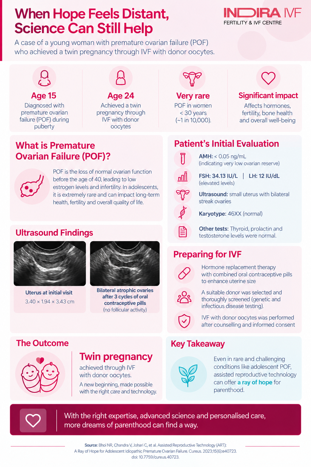
A published case report from Indira IVF documents how a woman with adolescent premature ovarian failure, a rare condition in which the ovaries stop functioning before reproductive life has fully begun, achieved a successful twin pregnancy through in vitro fertilization using donor eggs.
Based on: Bhoi NR, Chandra V, Johari C, Murdia K. "Assisted Reproductive Technology (ART): A Ray of Hope for Adolescent Idiopathic Premature Ovarian Failure." Cureus, 2023;15(6):e40723. Case managed at Indira IVF Hospital, India.
Table of Contents
- Introduction
- What is premature ovarian failure, and why is this case notable?
- Key facts at a glance
- The diagnostic picture
- Preparing the body for pregnancy
- The donor-egg IVF process
- Pregnancy and delivery outcome
- What causes idiopathic POI, and what are the treatment options?
- What this means for patients and families
Introduction
Premature ovarian failure in an adolescent is uncommon enough that most gynecologists will see only a handful of cases in an entire career. When it occurs with no identifiable cause, termed idiopathic, it can leave a young patient and her family without clear answers for years. This case report follows one such patient from her initial diagnosis at age 15 through to a successful pregnancy and delivery at 24, using in vitro fertilization (IVF) with donor eggs. The authors present it as a structured account of what timely diagnosis, hormonal preparation, and assisted reproduction can achieve in a condition often accompanied by uncertainty and emotional distress.
1 in 10,000 estimated incidence in women under 30 | Age 15 at first diagnosis | Age 24 at successful conception via IVF | 36 weeks gestation, healthy twin delivery |
What is premature ovarian failure, and why is this case notable?
Premature ovarian failure, more formally called premature ovarian insufficiency (POI), is diagnosed when a woman's ovaries stop functioning normally before age 40, resulting in absent or irregular periods and abnormally low estrogen levels. In India, natural menopause typically occurs between ages 40 and 50, so POI represents a significant departure from the expected reproductive timeline. Population estimates suggest it affects roughly one in 10,000 women under 30, and it is rarer still when it develops during adolescence, before secondary sexual characteristics such as breast development and menstruation have even become established.
This case is notable because it documents the full arc of care: an adolescent diagnosis with no identifiable underlying cause (hence "idiopathic"), years of hormone replacement therapy to support normal development, and, ultimately, a donor-egg IVF pregnancy that resulted in healthy twins. Case reports of this kind are not designed to prove how often such treatment succeeds. Their value lies in showing clinicians and patients what a well-managed pathway can look like, and in reinforcing existing guidance on early diagnosis and referral.
Key facts at a glance
A summary of the clinical course described in the report:
- The patient presented at age 15 with primary amenorrhea (she had never menstruated) and underdeveloped secondary sexual characteristics, prompting further evaluation.
- Blood tests showed a markedly elevated FSH (34.13 IU/L) and an anti-Mullerian hormone (AMH) below 0.05 ng/mL, both consistent with a depleted ovarian reserve, alongside very low estrogen (20 pg/mL).
- Her karyotype was normal (46XX), ruling out common chromosomal causes such as Turner syndrome, and no genetic or family history explained the condition. It was therefore classified as idiopathic.
- Ultrasound imaging showed a small, underdeveloped uterus and bilateral streak ovaries (ovaries with essentially no follicles or eggs).
- She was placed on hormone replacement therapy, which supported normal pubertal development, and later underwent IVF with donor eggs and her husband's sperm at age 24.
- The pregnancy was a dichorionic diamniotic twin pregnancy (each twin with its own placenta and amniotic sac, generally the lower-risk twin pregnancy type), which progressed uneventfully through standard prenatal screening.
- She delivered healthy twins by cesarean section at 36 weeks of gestation.
The diagnostic picture
At her first documented visit, the patient showed no meaningful breast or pubic hair development (Tanner stage 1) and no axillary hair, alongside primary amenorrhea. Blood work ruled out several common alternative explanations: testosterone, DHEA, and prolactin levels were normal, and thyroid antibodies were absent. Her FSH and LH were elevated while estrogen was very low, a hormonal pattern that points specifically to the ovaries themselves rather than to the brain's signaling centers as the source of the problem. Her karyotype came back as a normal female pattern (46XX), and pedigree evaluation found no inherited condition in the family.
Table 1. Hormone and imaging findings at diagnosis
Test | Result | What it indicates |
|---|---|---|
FSH (follicle-stimulating hormone) | 34.13 IU/L | Elevated, suggesting the ovaries are not responding to normal hormonal signals |
LH (luteinizing hormone) | 12 IU/dL | Elevated, consistent with ovarian-level failure |
Estradiol (E2) | 20 pg/mL | Very low, indicating minimal ovarian hormone production |
AMH (anti-Mullerian hormone) | Below 0.05 ng/mL | Reflects an extremely depleted egg supply |
Karyotype | 46XX (normal female) | Rules out chromosomal causes such as Turner syndrome |
Ultrasound | Small uterus; bilateral streak ovaries | Confirms underdeveloped reproductive organs with minimal ovarian tissue activity |
The report notes that diagnosis of adolescent POI is frequently delayed, in part because there is no single, universally agreed set of diagnostic criteria for this age group. The authors point to a separate published case series of 17 adolescents with non-chromosomal, non-medication-related premature ovarian failure, in which the average age at diagnosis was 16.1 years; within that group, 58.8% presented with primary amenorrhea, 23.5% with secondary amenorrhea (periods that stopped after starting), and 17.6% with irregular, infrequent periods.
Preparing the body for pregnancy
Before fertility treatment could begin, the patient's uterus needed to reach a size capable of supporting a pregnancy, since it had not fully developed due to the lack of natural estrogen exposure during adolescence. She was started on combined oral contraceptive pills specifically to build up the uterine lining and increase uterine size. After three treatment cycles, her uterus had grown from 3.40 x 1.94 x 3.43 cm to 6.0 x 2.8 x 5.3 cm, with a healthy endometrial (uterine lining) thickness of 5.1 mm. A repeat ultrasound confirmed that both ovaries remained atrophic with no follicular activity, and a repeat AMH test came back below 0.01 ng/mL, effectively confirming that no eggs of her own were available.
The donor-egg IVF process
With no viable path to conception using her own eggs, the patient and her husband were counseled on the option of IVF using donor eggs, which they chose to pursue. A donor was selected and underwent standard screening for genetic conditions (including thalassemia) and infectious diseases (HIV, hepatitis B and C, and syphilis) before proceeding.
The donor underwent ovarian stimulation using a GnRH antagonist protocol, a standard IVF regimen. The husband's sperm, which had normal parameters, was used to fertilize the donor eggs via intracytoplasmic sperm injection (ICSI), a technique in which a single sperm is injected directly into an egg. Meanwhile, the patient's uterine lining was prepared using estradiol valerate to mimic the hormonal environment needed to support an embryo. Two embryos, both graded 5AA, a high-quality blastocyst grade, were transferred after laser-assisted hatching, a technique that thins the embryo's outer shell to support implantation.
Pregnancy and delivery outcome
Table 2. Pregnancy milestones
Stage | Finding |
|---|---|
Pregnancy test | Positive, 14 days after embryo transfer |
Ultrasound at 6 weeks | Dichorionic diamniotic twin pregnancy confirmed |
Nuchal translucency (12 weeks) | 2.4 mm and 2.2 mm, both within the normal range |
Aneuploidy screening (13 weeks) | Normal, via noninvasive prenatal testing |
Delivery | Healthy twins by cesarean section at 36 weeks |
Throughout the pregnancy, the patient also received consultations with a clinical psychologist as needed, reflecting a recognized aspect of care for patients with premature ovarian insufficiency: the diagnosis itself, and the path to parenthood it requires, can carry a significant emotional burden.
What causes idiopathic POI, and what are the treatment options?
"Idiopathic" means no specific cause could be identified. The report notes that one proposed mechanism involves disruption of the normal process by which egg cells are gradually lost through programmed cell death (apoptosis) over a woman's lifetime; in POI, this process may be accelerated, or a woman may simply be born with fewer eggs than usual, leading to an early depletion of ovarian reserve.
The report outlines several management options relevant to women diagnosed with POI, summarized below.
Table 3. Management options discussed for premature ovarian insufficiency
Option | Purpose |
|---|---|
Hormone replacement therapy | Replaces estrogen and progesterone the ovaries no longer produce; supports development, manages symptoms, and can improve fertility potential |
Donor-egg IVF | Recommended when a woman's own ovarian reserve is critically depleted, as in this case |
Gestational surrogacy | An option when a woman cannot safely carry a pregnancy herself, provided a male partner's sperm is usable |
Experimental approaches | Includes ovarian tissue transplantation and stem cell therapy, still considered investigational |
Psychological counseling | Supports patients through the emotional impact of diagnosis and treatment |
Lifestyle measures | Balanced diet, regular exercise, stress reduction, and avoiding smoking or excess alcohol, to support overall health |
The report also notes that spontaneous natural conception is possible in some women with POI, since ovarian function occasionally and unpredictably resumes, but the chance of this happening is estimated at only 5% to 10%, and there is currently no treatment that reliably restores natural fertility. This is why donor-egg IVF is generally presented as the most feasible and effective route to pregnancy once ovarian reserve has been confirmed as severely depleted.
What this means for patients and families
The central message of this case report is the value of early recognition. Premature ovarian insufficiency does not only affect fertility. It can also influence bone density, cardiovascular health, and psychological wellbeing, which is why the authors, along with broader clinical guidelines cited in the report, recommend prompt evaluation of adolescents who show delayed puberty or absent periods, followed by appropriate counseling for the patient and family regarding both health monitoring and future fertility options. For families navigating a similar diagnosis, this case illustrates that a structured pathway, hormone support to enable normal development, confirmation of ovarian reserve, and donor-egg IVF where indicated, has resulted in a successful pregnancy and healthy delivery. It should be read as an illustrative account of one patient's outcome rather than a guarantee of results for every case, since individual circumstances vary considerably.