Overview
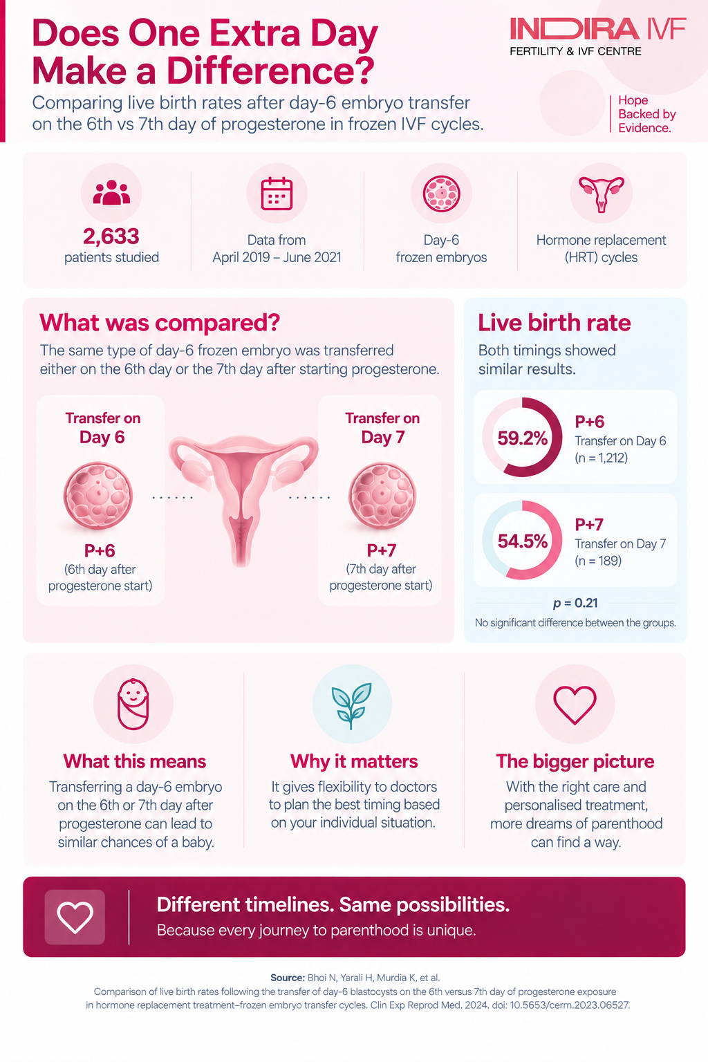
A large Indira IVF study of 2,633 patients examined whether slower-growing, day-6 blastocysts achieve better live birth rates when transferred on the 6th or the 7th day of progesterone exposure in hormone replacement FET cycles. The two timings produced comparable live birth rates, while three other factors emerged as the real predictors of success.
Table of Contents
- Introduction
- What is HRT-FET, and why does progesterone timing matter for day-6 blastocysts?
- Key findings at a glance
- How the study was conducted
- Results: pregnancy and live birth rates
- What actually predicts a live birth, regardless of timing?
- How this compares with earlier research
- Limitations worth knowing
- What this means for patients
Introduction
In frozen embryo transfer (FET) cycles prepared with hormone replacement therapy, timing is everything. The embryo and the uterine lining must reach a state of mutual readiness at precisely the same moment, a window that is carefully calibrated by the number of days of progesterone exposure before transfer. Most clinics transfer day-6 blastocysts, which grew a day more slowly in the laboratory, on the same schedule used for day-5 embryos: the 6th day of progesterone exposure. This large retrospective study asked a targeted question: would waiting one extra day, transferring on the 7th day instead, better match a slower-growing embryo to a more favourably timed uterine lining, and improve the chances of a live birth?
2,633 patients in the initial study cohort | 1,401 patients in the final matched analysis | 59.2% vs. 54.5% live birth rate, day 6 vs. day 7 (not statistically different) | 3 factors found to independently predict live birth |
What is HRT-FET, and why does progesterone timing matter for day-6 blastocysts?
In a hormone replacement treatment (HRT) frozen embryo transfer cycle, there is no corpus luteum (the temporary hormone-producing structure that forms after ovulation), so all of the progesterone that prepares the uterus for pregnancy must be supplied externally. The number of days of progesterone exposure before transfer is what determines the timing of the endometrial “window of implantation,” the narrow period during which the uterine lining is receptive to an embryo. Standard practice transfers day-5 blastocysts, and, by extension, day-6 blastocysts as well, on the 6th day of progesterone exposure (referred to in this study as P+6).
Day-6 blastocysts are embryos that took one extra day to reach the blastocyst stage in the laboratory, a sign that their development is somewhat slower than that of day-5 embryos. Because of this, researchers have questioned whether transferring these slower-growing embryos on the same P+6 schedule created a mismatch, and whether delaying transfer by one additional day, to P+7, might better align the embryo's developmental stage with the uterine lining's readiness. Only one earlier, smaller study had directly examined this question before now, and its results pointed toward a possible benefit for the 7th day, making this larger study a meaningful test of that finding.
Key findings at a glance
A summary of what this large retrospective analysis found:
- The study drew on 2,633 patients who underwent day-6 blastocyst transfer in HRT-FET cycles across 85 Indira IVF centres in India between April 2019 and July 2021.
- After statistical matching to ensure fair comparison, the final analysis included 1,212 patients transferred on the 6th day of progesterone (P+6) and 189 patients transferred on the 7th day (P+7).
- The live birth rate was 59.2% for P+6 versus 54.5% for P+7, a difference that did not reach statistical significance.
- The clinical pregnancy rate was 70.5% for P+6 versus 63.0% for P+7, a difference that was statistically significant, though it did not carry through to a significant difference in live births.
- Miscarriage rates were comparable between the two groups, at 15.9% for P+6 and 13.4% for P+7.
- Three factors emerged as independent predictors of live birth: the number of previous IVF attempts, the number of blastocysts transferred, and the endometrial thickness measured on the day progesterone treatment began. The timing of progesterone exposure itself, P+6 versus P+7, was not among them.
How the study was conducted
This was a retrospective cohort study, meaning researchers analyzed existing patient records rather than assigning patients to a treatment in advance. Data was drawn from the central registry of Indira IVF Hospital, covering 62,227 frozen embryo transfer cycles conducted between April 2019 and July 2021, from which 2,633 day-6 blastocyst transfer cycles using the HRT-FET protocol were identified.
To ensure a fair comparison between the P+6 and P+7 groups, despite this not being a randomized trial, the researchers used a statistical technique called propensity score matching. This method pairs patients from each group who have similar underlying characteristics, such as age, body mass index, ovarian reserve (measured by AMH), and endometrial thickness, so that any difference in outcomes is more likely to reflect the actual variable being studied (the timing of progesterone exposure) rather than pre-existing differences between the groups. After this matching process, 1,401 patients remained in the final analysis: 1,212 in the P+6 group and 189 in the P+7 group.
Table 1. Baseline characteristics of the matched study groups
Characteristic | Day 6 (P+6), n = 1,212 | Day 7 (P+7), n = 189 |
|---|---|---|
Female age at transfer | 31.10 ± 4.41 years | 31.61 ± 4.47 years |
Body mass index | 25.05 ± 4.01 kg/m2 | 24.93 ± 4.08 kg/m2 |
AMH level | 3.34 ± 2.59 ng/mL | 2.92 ± 2.48 ng/mL |
No previous IVF attempts | 53.6% of patients | 66.7% of patients |
Underwent PGT-A (genetic testing) | 3.1% of patients | 6.3% of patients |
Every patient in the study received oral estradiol valerate to prepare the endometrium, followed by intramuscular progesterone at a standard daily dose once the lining reached at least 7 mm in thickness. The only variable that differed by design was whether the day-6 blastocyst was transferred on the 6th or 7th day after progesterone began.
Results: pregnancy and live birth rates
The primary question the study set out to answer was whether live birth rate differed meaningfully between the two transfer timings. It did not.
Table 2. Reproductive outcomes, day 6 versus day 7 transfer
Outcome | Day 6 (P+6) | Day 7 (P+7) | Statistically significant? |
|---|---|---|---|
Positive pregnancy test rate | 72.6% | 66.1% | No (p = 0.066) |
Biochemical loss rate | 3.0% | 4.8% | No (p = 0.271) |
Clinical pregnancy rate | 70.5% | 63.0% | Yes (p = 0.037) |
Clinical miscarriage rate | 15.9% | 13.4% | No (p = 0.485) |
Live birth rate | 59.2% | 54.5% | No (p = 0.218) |
Multiple live birth rate | 16.5% | 14.8% | No (p = 0.559) |
Why the clinical pregnancy rate difference matters less than it appears: Although clinical pregnancy rate was statistically higher in the P+6 group, this difference did not persist through to live birth, the outcome that matters most to patients. This is a useful reminder that an early-stage pregnancy marker does not always predict the final result, and live birth rate remains the most meaningful measure of success.
What actually predicts a live birth, regardless of timing?
Rather than the timing of progesterone exposure, a multivariate regression analysis, a statistical method that isolates the independent effect of each factor while accounting for the others, identified three variables that meaningfully predicted whether a cycle ended in a live birth.
Table 3. Independent predictors of live birth
Predictor | Odds ratio | 95% confidence interval |
|---|---|---|
Number of previous IVF attempts | 3.246 | 2.429 to 4.337 |
Number of blastocysts transferred (two vs. one) | 2.011 | 1.461 to 2.768 |
Endometrial thickness on the day progesterone began | 1.143 | 1.022 to 1.28 |
In practical terms, a patient's prior IVF history, the decision to transfer one versus two embryos, and how well the uterine lining had developed by the time progesterone began were all more strongly associated with the chance of a live birth than whether the embryo was transferred on the 6th or 7th day. Female age and other embryo quality factors also showed expected associations with outcome in a related univariate comparison, though the timing of progesterone exposure did not.
How this compares with earlier research
Only one prior study, a smaller retrospective analysis by Roelens and colleagues published in 2020, had directly compared day-6 versus day-7 progesterone timing for day-6 blastocyst transfers. That earlier study reported a live birth rate that was numerically higher, though not statistically significant, on day 7 compared to day 6 (35.5% versus 21.5%), an effect the authors attributed mainly to a lower miscarriage rate on day 7.
The present study, with a substantially larger sample size, did not replicate that pattern: live birth rates were statistically comparable between the two timings, and miscarriage rates did not differ meaningfully either. The authors note that differences in sample size and in the route of progesterone administration (given by injection in this study, compared with a vaginal route in the earlier one) may help explain why the two studies reached different conclusions. A separate line of research using cleavage-stage (day-3) embryos, and another comparing day-5 and day-7 blastocyst transfers, has similarly found no statistically significant advantage to delaying transfer by a day or two, broadly consistent with the findings reported here.
Limitations worth knowing
Several factors are worth keeping in mind when interpreting these results:
- This was a retrospective study using existing patient records rather than a randomized controlled trial, meaning that despite statistical matching, some unmeasured differences between the groups could still have influenced the results.
- The P+7 group was considerably smaller than the P+6 group after matching (189 versus 1,212 patients), since transferring on the 7th day is a far less common clinical practice, which limits the statistical power available to detect smaller differences.
- A substantial proportion of patients who tested positive for pregnancy were lost to follow-up before live birth data could be recorded (46.0% in the P+6 group and 45.7% in the P+7 group), which may have introduced some selection bias into the final results.
- Most transfers in this study involved two blastocysts rather than one, which may limit how well these findings apply to clinics that predominantly perform single embryo transfers.
- The findings reflect data from a single hospital network within a defined time period and country, so results elsewhere may vary depending on local protocols and patient populations.
What this means for patients
For patients whose embryo took an extra day to reach the blastocyst stage, this study offers reassurance: the data suggest that transferring a day-6 blastocyst on the standard 6th day of progesterone exposure produces live birth outcomes comparable to delaying the transfer by an additional day. There does not appear to be a meaningful advantage to postponing transfer specifically to accommodate a slower-growing embryo. Instead, the factors shown here to matter most, a patient's treatment history, whether one or two embryos are transferred, and how well the uterine lining has developed, are the considerations worth discussing in detail with a fertility specialist when planning a frozen embryo transfer cycle.