
Overview
An endometrial biopsy examines the uterine lining and is usually advised for unusual bleeding, irregular periods, postmenopausal spotting, or a thickened lining on scans, and sometimes during infertility evaluation. Waiting for results can feel stressful, and pathology terms often sound scarier than they are. Some reports simply reflect hormonal changes, while others show inflammation, polyps, or cells that need monitoring. How a result is interpreted depends on factors like age, menstrual history, and menopausal status. This article breaks down what biopsy results commonly mean, which findings are normal, what abnormal terms indicate, and when further follow-up may be needed.
Table of Contents
- Why Is an Endometrial Biopsy Done?
- How Does the Endometrial Biopsy Procedure Happen?
- How Long Do Biopsy Results Take?
- What Do Normal Biopsy Results Mean?
- What is Endometrial Hyperplasia?
- Do Biopsy Results Always Mean Cancer?
- What Does Atypia Mean in a Biopsy?
- Can Infections Appear in Biopsy Findings?
- Why Do Some Reports Say Insufficient Tissue?
- What happens after abnormal results?
- Does Menopause Affect Biopsy Interpretation?
- When is a repeat biopsy advised?
- Conclusion
Why Is an Endometrial Biopsy Done?
The biopsy helps doctors study the cells lining the uterus.
Usually, the test is recommended when bleeding patterns change unexpectedly. Some women notice heavier periods than before. Others develop spotting between cycles. In postmenopausal patients, any slight bleeding must be investigated since bleeding following menopause is never considered to be a normal occurrence.
An endometrial biopsy may be recommended in cases where there is:
- Excessive menstrual bleeding
- Irregular cycles
- Post-menopausal bleeding
- Thickened endometrium by ultrasound
- Chronic hormonal imbalance
- Issues concerning fertility
- Abnormalities of the uterus
Scans alone do not always offer an explanation for symptoms experienced. This is where a biopsy is helpful since it reveals the actual condition of the endometrial lining.
How Does the Endometrial Biopsy Procedure Happen?
Before the Procedure
- History will generally be taken concerning the patient’s menstrual background, unusual vaginal bleeding, current medications, and past gynaecological issues.
- Ultrasonography may have demonstrated endometrial thickening or other findings necessitating further testing.
- Generally, women are advised to notify the doctor if there is a chance of pregnancy, if they take any blood-thinning medication, and if they suffer from any allergies or infections.
- In some cases, the doctor may suggest taking a mild pain-relieving medicine before the procedure to reduce cramping.
- Most endometrial biopsies are done in the outpatient clinic, so hospital admission is usually not required.
- General anaesthesia is not commonly needed because the procedure is usually short.
During the Procedure
- The woman lies on the examination table, similar to a routine pelvic examination.
- A speculum is placed inside the vagina so the doctor can view the cervix properly.
- A narrow tube is placed through the cervix so the doctor can reach the lining inside the uterus.
- Next, a small instrument is used to collect tissue from the uterine lining.
- The procedure is usually done while the woman remains awake.
- Some women feel cramps during the sampling part of the procedure, often similar to strong menstrual cramps.
- Others mainly notice pressure in the pelvis or a short-lasting, sharp pain that settles quickly once the sample is taken.
- The overall experience differs quite a bit from one woman to another.
- After the sample is collected, it is sent to the laboratory where the cells are examined under a microscope.
After the Procedure
- Light spotting after the biopsy is fairly common.
- Some women continue feeling mild cramps or lower abdominal discomfort for a few hours afterwards.
- A few women may also notice temporary lower back pain.
- These symptoms usually go away without treatment.
- Depending on the situation, the physician might recommend avoiding sexual intercourse and tampons for some time after the procedure.
- Women are generally advised to contact their doctor if they develop fever, severe pain, heavy bleeding, or foul-smelling discharge afterwards.
- In many cases, the biopsy report becomes available within a few days, although certain reports may take longer if additional testing is needed.
How Long Do Biopsy Results Take?
Sometimes turnaround time varies significantly by laboratory and institution; the results arrive earlier, while certain cases may take longer if additional microscopic examination or review is required.
Waiting for the report can feel stressful for many women, especially when abnormal bleeding has already been present for some time. The final interpretation of the biopsy usually depends not only on the laboratory findings but also on symptoms, ultrasound appearance, age, menstrual history, and menopausal status.
What Do Normal Biopsy Results Mean?
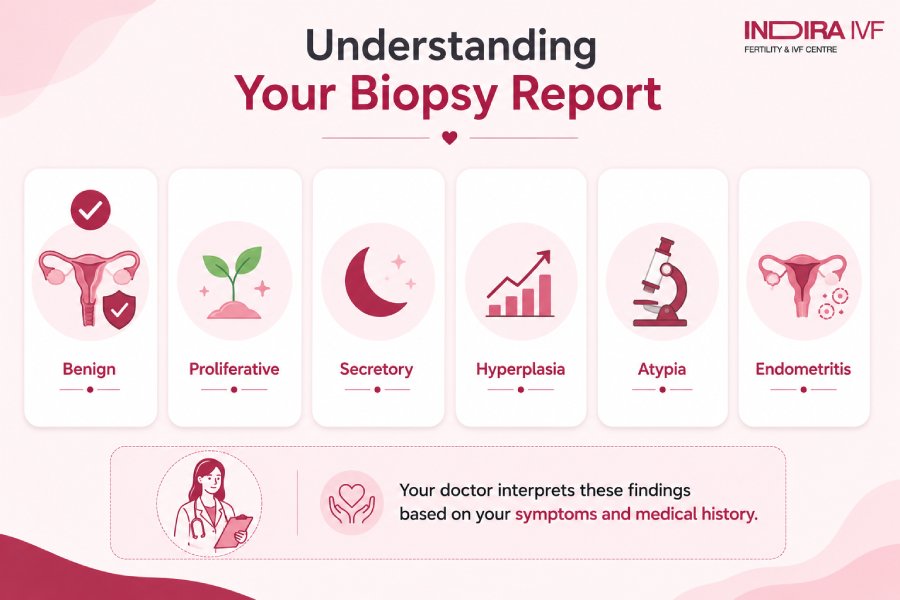
Many endometrial biopsy reports eventually turn out to be normal. A normal report generally means that no dangerous or cancer-related abnormality was identified in the tissue sample taken from the uterine lining.
Biopsy Term | What It Usually Means |
Benign endometrium | No cancer-related changes seen |
Proliferative endometrium | Normal lining growth before ovulation |
Secretory endometrium | Normal post-ovulation changes |
No evidence of malignancy | No cancer was identified in the sample |
The appearance of the uterine lining naturally changes at different stages of the menstrual cycle, so doctors interpret the report alongside the woman’s symptoms and hormonal status.
What is Endometrial Hyperplasia?
Endometrial hyperplasia involves abnormal proliferation of the tissue layer inside the womb. This disorder usually gets detected through an endometrial biopsy that is taken on account of some unusual symptoms, such as heavy menstrual flow or bleeding at menopause.
In many women, the thickening develops because estrogen continues to stimulate the uterine lining without sufficient progesterone to balance it. The condition is seen more often in women with irregular ovulation, polycystic ovarian syndrome, obesity, diabetes, or hormonal changes occurring around menopause.
An endometrial biopsy helps determine the exact type of hyperplasia present.
Types commonly seen in biopsy reports:
- Hyperplasia without atypia
The lining appears thickened, but the cells do not show major abnormal changes under the microscope.
- Atypical hyperplasia
The cells begin showing abnormal microscopic features. This finding is monitored more carefully because the risk of developing endometrial cancer becomes higher compared to simple hyperplasia.
The treatment will depend on various factors like age, symptoms, menopause, and any future plans of becoming pregnant. Some people can benefit from hormonal treatment, while others need surgery.
Do Biopsy Results Always Mean Cancer?
No. Most women undergoing an endometrial biopsy do not have cancer. Abnormal bleeding may happen because of:
- Hormonal imbalance
- Fibroids
- Polyps
- Irregular ovulation
- Benign thickening of the uterine lining
Still, biopsy remains important because it helps rule out serious disease.
In some biopsy reports, the laboratory may identify cancerous changes in the uterine lining. In certain cases, the biopsy report may mention cancer-related findings such as endometrial carcinoma, adenocarcinoma, or malignant changes involving the uterine lining.
At this point, the healthcare provider will recommend conducting additional tests to gauge the extent of the disorder and plan subsequent treatment.
What Does Atypia Mean in a Biopsy?
Sometimes, the biopsy report may mention the word “atypia.” This means certain cells in the uterine lining do not appear completely normal under microscopic examination.
It does not always mean cancer is already present. However, doctors generally pay closer attention to these changes because some abnormal cells may gradually develop further changes over time if they are not treated or monitored properly.
After such findings, some women may need:
- Repeat the biopsy after some time
- Hormonal treatment
- Regular follow-up visits
- Surgery in selected situations
The next step is usually decided after considering the woman’s age, symptoms, biopsy findings, and whether menopause has already occurred.
Can Infections Appear in Biopsy Findings?
Yes. Occasionally.
Some biopsy reports show inflammation inside the uterine lining.
Terms that may appear include:
- Endometritis
- Chronic inflammation
- Plasma cells
Some women with infection-related changes may develop:
- Pelvic pain
- Fever
- Irregular bleeding
- Vaginal discharge
Others may not notice obvious symptoms at all.
In fertility evaluation, chronic inflammation sometimes receives more attention because persistent inflammation may interfere with implantation. Treatment may involve antibiotics depending on the underlying cause.
Why Do Some Reports Say Insufficient Tissue?
There are cases in which the specimen obtained is too small for an accurate assessment.
This can be seen in:
- Post-menopausal patients
- Those with a thin endometrium
- Challenging sample collection techniques
Some terms used in the report include:
- Insufficient tissue
- Small specimen
- Scanty specimen
- Inadequate tissue
It doesn’t mean that a cancer diagnosis will follow automatically.
Still, if symptoms continue, doctors may advise additional evaluation, such as:
- Repeat biopsy
- Hysteroscopy
- Dilation and curettage
- Repeat ultrasound
Persistent symptoms usually matter more than one isolated report line.
What happens after abnormal results?
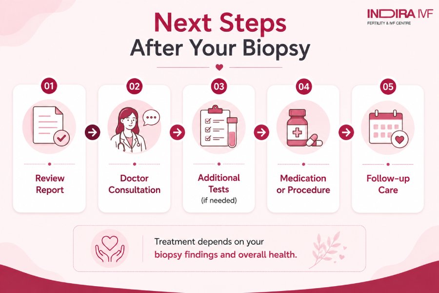
That depends entirely on what the biopsy shows.
Some women only require monitoring and follow-up scans.
Others may need:
- Hormonal medication
- Repeat biopsy
- Removal of polyps
- Hysteroscopy
- Surgery
- Cancer treatment
There is no single treatment plan that fits everybody.
Doctors usually combine the following before deciding the next step:
- Pathology findings
- Symptoms
- Ultrasound appearance
- Age
- Menopausal status
- Medical history
Does Menopause Affect Biopsy Interpretation?
Yes. Menopause causes thinning of the endometrium (uterine lining). It means that the occurrence of any vaginal bleeding in women after menopause requires immediate investigation.
These examinations include an ultrasound, biopsy, and/or hysteroscopy. A thickened endometrial lining after menopause can be worrying due to conditions such as hyperplasia, polyps, or cancerous growths, although most women are often diagnosed with harmless conditions.
Common Terms Seen in Biopsy Reports
Report Term | Meaning |
Benign endometrium | Noncancerous tissue |
Proliferative endometrium | Normal estrogen-related growth |
Secretory endometrium | Normal post-ovulation pattern |
Hyperplasia | Thickening of the uterine lining |
Atypia | Abnormal cellular appearance |
Atrophic endometrium | Thin lining is often seen after menopause |
Endometritis | Inflammation or infection |
Malignancy | Cancerous cells present |
Pathology reports often sound more frightening than they actually are.
That is why doctors explain the report alongside the overall clinical picture rather than relying solely on a single medical term.
When is a repeat biopsy advised?
Doctors may advise repeat testing if:
- Bleeding continues
- Ultrasound findings remain abnormal
- The tissue sample was insufficient
- Hyperplasia needs monitoring
- Symptoms worsen over time
Women with higher risk factors sometimes need closer follow-up.
These risk factors may include:
- Obesity
- Diabetes
- Long-term estrogen exposure
- Persistent postmenopausal bleeding
- Family history of uterine cancer
Follow-up plans are usually individualised.
Conclusion
Endometrial biopsy results are used to examine changes affecting the lining of the uterus. The report may show normal hormonal patterns, inflammation, thickening of the lining, hyperplasia, abnormal cellular changes, or, in some cases, cancer-related findings.
However, many of these women often end up being diagnosed with non-cancerous disorders. Irregularities in hormone levels, ovulation, the presence of fibroids, and even benign endometrial thickening are common causes for abnormal bleeding.
The biopsy report is not interpreted in isolation. Doctors usually assess it alongside symptoms, ultrasound findings, menstrual history, age, and menopausal status before deciding on the next step in management.
Understanding the meaning of the report often helps women approach treatment decisions with better clarity and less unnecessary anxiety.