
Overview
The journey from a single fertilised egg to a fully developed living organism starts during embryonic development. The process involves coordinated cellular processes that are considered the foundation for life. Among these stages, one of the most important ones is blastocyst formation, development, and implantation. This is because they mark the stage when the embryo moves from floating freely to attaching itself to the uterine lining, which is the beginning of pregnancy.
A blastocyst is a ball of cells that is formed during early pregnancy (about 5-6 days) after a sperm fertilises an egg. This article explains what a blastocyst is, the formation of a blastocyst, its purpose, and other important elements.
Table of Contents
- What is a blastocyst?
- What Does a Blastocyst Look Like?
- What is the Significance of the blastocyst?
- What are the Stages of Blastocyst Formation?
- What happens after a blastocyst is formed?
- Are There Any Signs of Blastocyst Formation?
- When Blastocyst Formation Fails?
- What’s the Difference Between a Blastocyst and an Embryo?
- Conclusion
What is a blastocyst?
A blastocyst is an early stage of embryonic development. In this, the fertilised egg (zygote) undergoes a complete event of cell division while travelling through the fallopian tube. It is a cluster of cells that forms about 5-6 days after conception through a process called blastulation. During cell division, the embryo changes from a single cell to a compact cluster of cells called the morula, which transforms into the blastocyst.
The blastocyst is an important stage in pregnancy, during which the embryo must develop to be implanted successfully. Cells in the blastocyst multiply and separate into two types. The inner cell mass develops into the fetus, while the outer trophoblast cells contribute to placental development.
The blastocyst stage is an important stage in the IVF (in vitro fertilisation). During IVF, the egg is fertilised in a culture dish and allowed to develop into a blastocyst in the lab. Its growth and development are monitored. Doctors grade them based on their maturity, appearance, and cell count.
What Does a Blastocyst Look Like?
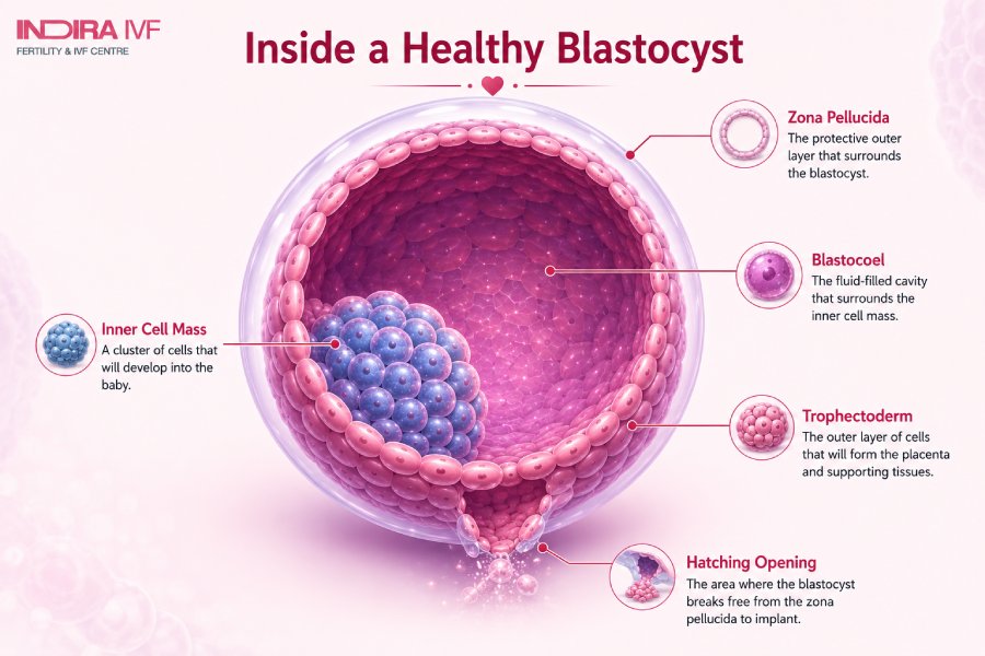
A blastocyst looks like a hollow ball of cells (spherical in shape), and has -
- A clear outer protective layer called the zona pellucida
- A ball of cells called trophoblasts that are under the zona pellucida
- An inner cluster of cells attached to one of the trophoblasts, known as the inner cell mass
- Fluid cavity inside the blastocyst, known as the blastocele
The size of the blastocyst measures about 0.1 mm to 0.2mm in diameter, and contains approximately 200-300 cells. Blastocysts have different sizes and appearances. During IVF, doctors identify a high-quality blastocyst with a clear inner cell mass and tight trophectoderm.
Also, the size of the blastocyst helps healthcare professionals understand how well it has developed. Moreover, it helps determine if it is ready to be transferred to the uterus.
Cell Specification in the Blastocyst
As the blastocyst continues to develop, its cells begin to organise into specialised cell groups.
- The trophoblast, which forms the placenta and helps with implantation.
- The epiblast gives rise to the fetus (baby).
- And, thePrimitive Endoderm helps form supportive tissues like the yolk sac to support early development.
Apart from supporting the embryo’s growth, each group has a specific role. The embryo controls these changes through several biological processes. It does so by -
- Turning certain genes on and off (gene expression)
- Communicating between the cells (cell signalling)
- Also, communicating with the neighbouring cells (cell-to-cell contact)
As the inner cell mass (ICM) grows inside the blastocyst, its cells start splitting into two groups - the epiblast and primitive endoderm. The split is monitored by a signalling molecule known as fibroblast growth factor (FGF). The FGF sends a signal through the MAP kinase pathway. It is a pathway that helps control the cell behaviour and its development.
What is the Significance of the blastocyst?
The blastocyst stage is a very important part of embryonic and fetal development. This is because, if the blastocyst does not implant in the lining of the uterus, pregnancy is not possible. For implantation to occur, hormones trigger a process called hatching, which occurs 1-3 days after a blastocyst is transferred to the uterus. During hatching, the blastocyst sheds its clear outer membrane.
The outer cell (trophectoderm) attaches to the lining of the uterus by releasing a sticky protein that helps it bind to the endometrium. The outer cell transforms into a placenta, which carries oxygen and nutrients to the developing fetus.
Extraembryonic tissues contribute to the formation of the amniotic sac, which is a clear fluid that surrounds and protects the baby. Whereas the ICM (inner cell mass) of the blastocyst continues to grow, it develops into a fetus.
What are the Stages of Blastocyst Formation?
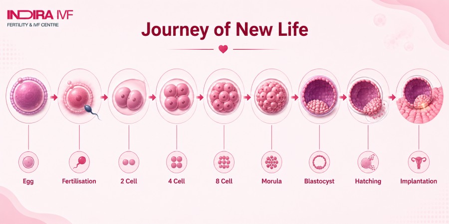
Blastocyst formation occurs in the fallopian tube during natural pregnancy, where the fertilised egg divides as it travels to the uterus. Here’s how a blastocyst forms:
Fertilisation to Early Embryo
The process begins when the sperm fertilises the egg in the fallopian tube. Thus, forming a single-celled zygote, which undergoes rapid cell division, a process called cleavage. During the division process, there is no growth, so the cells continue to diminish in size.
The zygote divides into two, then four, and by day 3, the cells narrow down to eight. These are called blastomeres. By day 4, the embryo develops into a morula, which is a solid mass of 16-32 cells. The size of the morula is like a tiny mulberry.
Cell Differentiation and Organisation
Next, the morula undergoes a transformation when its cells begin to differentiate and organise. One of the most important events that takes place is called “compaction.” During this, the cells move closer together and stick to one another. The process makes the embryo look like a smooth, compact ball. It takes place when the outer cell forms connections, called tight and gap junctions, which help the cells attach and communicate with each other.
Next, the cells start to polarise, during which the inner and outer surfaces are developed. Some cells stay outside, while others remain inside the embryo. Then, fluid begins to collect within the compacted morula, a process called cavitation. It creates a central, fluid-filled cavity in the embryo, called the blastocoel. The blastocoel expands, assisted by active transport of sodium ions into the intercellular spaces, followed by water by osmosis.
As the blastocoel expands, the cells separate into two main groups, such as -
- Outer cell layer, which later forms the trophoblast and contributes to the placenta
- Inner cell mass, which develops into the fetus
Once these features are formed completely, the embryo is called a blastocyst. This usually forms around 5-6 days after fertilisation.
What happens after a blastocyst is formed?
After the blastocyst has formed, here's what happens -
Hatching
As the blastocyst nears the endometrium, the zona pellucida breaks down. The blastocyst is released from the protective layer (the shell) so that it can interact with the uterus. This process is known as hatching. Here’s how hatching takes place -
Natural Hatching
In natural conception, the zona pellucida weakens as the embryo grows and the blastocyst expands. At the same time, the blastocyst releases enzymes, such as trypsin-like proteases, that soften and break down the shell.
IVF
In IVF, too, the embryo hatches naturally, but the entire process happens in a lab. Sometimes the zona pellucida may thicken or harden. The hard shell prevents it from hatching on its own. Hence, doctors may use assisted hatching. The most used method is laser-assisted hatching. This helps create a tiny opening in the zona pellucida using a laser before the embryo is transferred to the uterus.
Implantation
After the blastocyst hatches, it attaches to the uterine lining. The process occurs after 5-6 days of fertilisation. The trophectoderm cells attach to the uterine lining.
Confirmation of the Pregnancy
Once the implantation takes place, doctors test hCG levels either by urine or blood to confirm pregnancy. Furthermore, they verify the same using ultrasound.
Are There Any Signs of Blastocyst Formation?
The signs of blastocyst formation cannot be seen in a natural pregnancy since the process happens inside the body. However, when the egg is fertilised outside the womb (in the lab) during IVF, the formation of the inner cell mass and outer layer of trophoblast cells can be seen and monitored.
Also, the blastocyst itself does not produce any symptoms. However, as the pregnancy takes place, some people may later experience implantation-related symptoms, such as cramping,light spotting, etc. This mostly happens when the blastocyst is attached to the uterine lining.
When Blastocyst Formation Fails?
The formation of a blastocyst may fail if the developing embryo cannot continue normal growth during the early stages. Since the process depends on cell division, several factors can affect embryo development. Some of them are -
- Energy production
- Genetic activity
- Communication between cells
One of the major reasons for blastocyst formation failure is chromosomal aneuploidy. It means the embryo does not have the correct number of chromosomes. They might either be missing (monosomy) or have an extra copy (trisomy). These errors are incompatible with normal embryonic development.
Another reason may be poor egg or sperm quality. Whether through natural conception or IVF, healthy sperm and eggs are needed for fertilisation to occur. It may also fail due to -
- Hormonal imbalances
- Impaired energy production
- Inadequate mitochondrial function
- Unfavourable culture conditions during IVF
- The embryo may not receive a proper nutrient supply
Note: Failure of blastocysts does not mean that future pregnancies will fail.
What Happens When Blastocyst Formation Fails?
As the blastocyst does not form, the embryo's development stops, too. This is because it is unable to undergo the structural and metabolic changes required to develop into a blastocyst. This might result in the embryo stopping dividing and developing abnormally. It can also fail to form important structures, such as the blastocoel, the inner cell mass that forms the fetus, or the trophoblast layer.
What’s the Difference Between a Blastocyst and an Embryo?
Here are some key differences -
Blastocyst Vs. Embryo | |
A blastocyst is a thin-walled, hollow, ball-like structure that forms in the early stage of embryonic development. | An embryo is a rudimentary stage of a living being that shows potential for development. |
Develops from the zygote | Occurs from the ICM of the blastocyst |
Formed through a process called blastulation | Undergoes gastrulation, neurulation, and organogenesis |
Forms 4-5 days to two weeks after fertilisation | Forms 2-11 weeks after fertilisation |
Only found in mammals | Can be found in plants and animals |
Conclusion
Blastocyst formation is a process in which the zygote undergoes a series of cell divisions. The blastocyst consists of two distinct cell types: the inner cell mass (ICM) and the trophoblast. The formation of a blastocyst can either occur in the uterus or outside the uterus (in the fallopian tubes).
After the blastocyst forms, it descends into the uterus for hatching. During hatching, the outer shell of the embryo (zona pellucida) breaks as the blastocyst expands. This allows the embryo to attach to the uterine lining for a successful pregnancy.
The process of how a blastocyst is formed is the same for both natural pregnancy and IVF. However, the only difference is that during an assisted pregnancy, the process takes place in a lab, where it is monitored by IVF specialists.