
Overview
A case series from Indira IVF documents nine instances of heterotopic pregnancy, a rare condition in which an ectopic pregnancy and a normal pregnancy inside the uterus occur at the same time, following IVF and embryo transfer. Prompt diagnosis and surgery preserved the uterine pregnancy in every treated case, and eight of the nine pregnancies had resulted in live births by the time of publication.
Based on: Lunkad A, Patil B, Chandra V, Bhoi NR. "Pregnancy Outcomes of Heterotopic Pregnancy Following IVF-ET: A Case Series." Journal of Clinical Images and Medical Case Reports, 2024;5(8):3210. Conducted at Indira IVF Hospital, Pune, India.
Table of Contents
- What This Case Series Found About Heterotopic Pregnancy
- What is heterotopic pregnancy, and why does IVF raise the risk?
- Key facts at a glance
- How these nine cases were identified and treated
- Case-by-case outcomes
- Why heterotopic pregnancy is becoming more common with IVF
- The one case managed without surgery
- Limitations worth knowing
- Key Results at a Glance
- What this means for patients and fertility clinics
What This Case Series Found About Heterotopic Pregnancy
Study at a Glance | |
|---|---|
Item | Details |
Evidence type | Retrospective case series |
Cases identified | 9 cases of heterotopic pregnancy, out of 4,575 clinical pregnancies |
Study period | July 1, 2020 to June 30, 2023 |
Clinical setting | Indira IVF Hospital, Pune, following IVF and embryo transfer (5,936 ET cycles) |
Management | Laparoscopic surgery in 8 cases; potassium chloride injection and cervical cerclage in 1 cervical case |
Main outcome | 8 of 9 intrauterine pregnancies resulted in live births; 1 pregnancy was ongoing at the time of publication |
Publication | Journal of Clinical Images and Medical Case Reports, 2024 |
DOI | 10.52768/2766-7820/3210 |
Heterotopic pregnancy refers to the rare but serious situation where a pregnancy develops simultaneously inside and outside the uterus, most often as a normal pregnancy in the uterus alongside an ectopic pregnancy in a fallopian tube. In natural conception, this is extremely rare, but the risk rises meaningfully with IVF and embryo transfer, particularly when more than one embryo is placed during a cycle. This case series documents nine such cases identified at one fertility centre over three years, describing how each was diagnosed, how the ectopic pregnancy was managed, and what happened to the accompanying uterine pregnancy afterward.
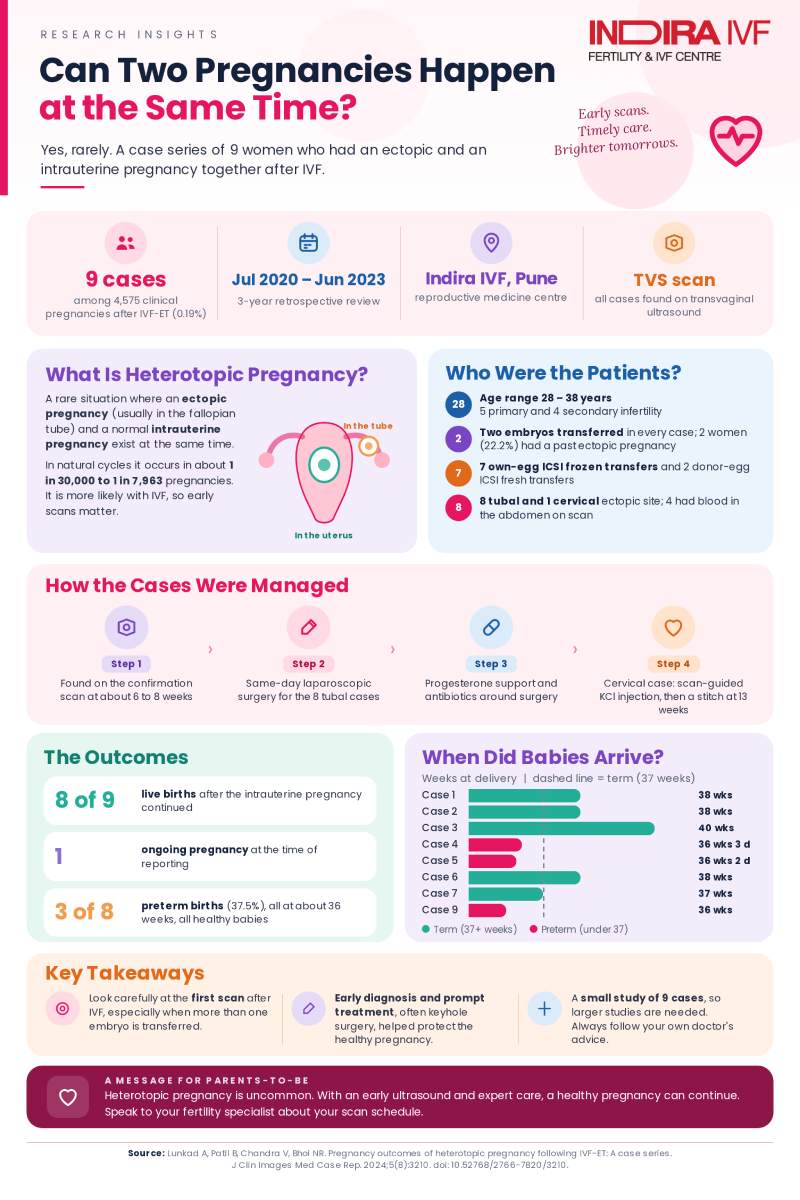
9 of 4,575 clinical pregnancies diagnosed with heterotopic pregnancy (0.19%) | 8 tubal, 1 cervical sites of the ectopic pregnancy across the nine cases | 8 of 9 pregnancies resulted in live births by the time of publication | 3 of 8 delivered pregnancies were preterm (37.5%) |
What is heterotopic pregnancy, and why does IVF raise the risk?
Heterotopic pregnancy is the simultaneous presence of both an ectopic pregnancy, most often located in a fallopian tube, and a normal, viable pregnancy inside the uterus. In natural, unassisted conception, this is exceptionally rare, occurring in roughly 1 in 30,000 to 1 in 7,963 pregnancies. With IVF and embryo transfer, however, the reported incidence in the wider medical literature can be as high as 1 in 100 pregnancies, a substantial increase attributed to factors such as the number of embryos transferred per cycle, pelvic inflammatory disease, prior ectopic pregnancies, and ovarian hyperstimulation syndrome.
At the fertility centre in this study, 5,936 embryo transfer cycles yielded 4,575 clinical pregnancies over the three-year study period, giving a clinical pregnancy rate of 77.07%. Among these, nine cases were identified as heterotopic pregnancy, an incidence of 1.96 per 1,000 clinical pregnancies, or 0.19%, following IVF and embryo transfer.
Key facts at a glance
A summary of the case series as a whole:
- Nine cases of heterotopic pregnancy were identified out of 4,575 clinical pregnancies at one fertility centre between July 2020 and June 2023.
- Eight of the nine cases involved a tubal ectopic pregnancy, while one case involved a rare cervical ectopic pregnancy.
- Maternal age ranged from 28 to 38 years; five patients had primary infertility and four had secondary infertility.
- Two patients (22.2%) had a history of a previous ectopic pregnancy, a recognized risk factor for heterotopic pregnancy.
- Seven patients conceived using their own eggs through frozen embryo transfer, while two patients with poor ovarian reserve used donor eggs with a fresh embryo transfer.
- Hemoperitoneum (blood in the abdominal cavity, a sign of internal bleeding) was detected by ultrasound in four of the nine cases.
- Eight patients underwent laparoscopic surgery to remove the ectopic pregnancy on the same day it was diagnosed, and all had their intrauterine pregnancy successfully preserved.
- Among the eight pregnancies that had delivered by the time of publication, all resulted in live births, though three (37.5%) were preterm; one pregnancy remained ongoing at the time of publication.
How these nine cases were identified and treated
All nine cases were identified through transvaginal sonography (TVS) during routine confirmation scans, typically between 6 and 8 weeks of gestation. Before surgery, all patients received intramuscular progesterone (100 mg), supplemented postoperatively by vaginal progesterone gel and dydrogesterone, alongside antibiotic therapy, to support the ongoing intrauterine pregnancy.
For the eight cases with a tubal or tubal-stump ectopic pregnancy, laparoscopy and salpingectomy (or, in one case, removal of the ectopic mass alone) were the chosen management strategy, performed the same day as diagnosis. In one instance, a patient who had previously undergone bilateral salpingectomy for an earlier ectopic pregnancy was found to have the new ectopic pregnancy implanted in the retained tubal stump, confirmed on histopathological examination. Laparoscopic surgery at this centre was performed under general intravenous anesthesia, with carbon dioxide pressure kept below 12 mmHg and the patient positioned in a dorsal supine position, an approach the authors note has been shown to be safe for both mother and baby.
Table 1. Summary of the nine heterotopic pregnancy cases
Case | Age | Infertility type | Gestational age at detection | Management | Ectopic site | Outcome |
|---|---|---|---|---|---|---|
1 | 28 | Secondary | 6 weeks 6 days | Laparoscopic ectopic mass removal | Tubal stump | Live birth, 38 weeks |
2 | 31 | Secondary | 6 weeks 4 days | Laparoscopic left salpingectomy | Tubal | Live birth, 38 weeks |
3 | 32 | Primary | 6 weeks 2 days | Laparoscopic left salpingectomy | Tubal | Live birth, 40 weeks |
4 | 33 | Primary | 8 weeks | Laparoscopic right salpingectomy | Tubal | Live birth, 36 weeks 3 days |
5 | 30 | Secondary | 7 weeks 1 day | Laparoscopic right salpingectomy with bilateral tubal clipping | Tubal | Live birth, 36 weeks 2 days |
6 | 33 | Primary | 6 weeks 4 days | Laparoscopic right salpingectomy | Tubal | Live birth, 38 weeks |
7 | 33 | Secondary | 6 weeks 4 days | Laparoscopic left salpingectomy | Tubal | Live birth, 37 weeks |
8 | 36 | Primary | 6 weeks 2 days | Laparoscopic left salpingectomy | Tubal | Ongoing pregnancy at publication |
9 | 38 | Primary | 6 weeks 4 days | Intracervical potassium chloride injection under ultrasound guidance | Cervical | Live birth, 36 weeks |
Case-by-case outcomes
Seven of the eight surgically managed pregnancies reached term or near-term delivery (37 to 40 weeks), while two (cases 4 and 5) were mildly preterm at 36 weeks. The ninth case, managed without surgery, also delivered preterm at 36 weeks. Across the eight pregnancies that had delivered by the time of publication, every one resulted in a live birth, and one further pregnancy (case 8) remained ongoing at the time the case series was written.
For comparison, the authors cite a separate published series of 65 heterotopic pregnancy cases from a single institution, which reported a 93.8% live birth rate, with surgical management used in 84.6% of cases, figures broadly consistent with the outcomes seen in this smaller series.
Why heterotopic pregnancy is becoming more common with IVF
The authors note that the rise in heterotopic pregnancy cases correlates directly with the number of embryos transferred per embryo transfer cycle. Several mechanical factors during the embryo transfer procedure are thought to contribute, including misplacement of the catheter tip, excess culture medium, pressure applied during embryo injection, and endometrial bleeding, any of which might allow an embryo to implant in the fallopian tube rather than, or in addition to, the uterus.
Why early ultrasound matters: High-resolution transvaginal ultrasound is described as the preferred, cost-effective method for diagnosing heterotopic pregnancy. The authors note that routine transvaginal ultrasound around day 27 after embryo transfer can help catch heterotopic pregnancy early, reducing missed diagnoses. At this centre, a comparatively lower incidence of heterotopic pregnancy was observed, which the authors suggest may be partly attributable to a preference for blastocyst-stage frozen embryo transfer, which made up around 65% of frozen transfer cycles, since blastocyst-stage transfer has been associated with a lower rate of ectopic and heterotopic pregnancy than cleavage-stage transfer in other studies.
The one case managed without surgery
The ninth case in this series, involving a cervical ectopic pregnancy alongside a viable intrauterine pregnancy, was managed differently from the other eight. Rather than surgery, the care team used an intracervical potassium chloride injection under ultrasound guidance to address the ectopic pregnancy, followed by serial weekly ultrasounds to monitor the cervix. These follow-up scans showed the ectopic sac gradually resorbing, while the intrauterine pregnancy remained viable. At 13 weeks of gestation, a cervical cerclage (a stitch placed around the cervix to provide additional support) was performed. The intrauterine pregnancy then progressed normally, culminating in a preterm delivery at 36 weeks that resulted in a healthy baby despite the preterm labor.
Limitations worth knowing
Several factors are worth keeping in mind when interpreting this case series:
- This is a small, retrospective case series of nine patients from a single fertility centre, which limits how precisely the findings can be generalized to other clinical settings or larger patient populations.
- The retrospective nature of the study made it difficult to precisely pinpoint the exact location of each tubal ectopic pregnancy, such as whether it occurred in the ampullary or interstitial portion of the fallopian tube.
- One of the nine pregnancies remained ongoing at the time of publication, meaning its final outcome was not yet known when this case series was written.
- As a single-centre report, the specific surgical techniques, protocols, and outcomes described may not directly apply to every fertility clinic or surgical team.
Key Results at a Glance
Measure | Result |
|---|---|
Incidence of heterotopic pregnancy at this centre | 9 of 4,575 clinical pregnancies (0.19%, or 1.96 per 1,000) |
Reported incidence in natural conception (cited literature) | Approximately 1 in 30,000 to 1 in 7,963 pregnancies |
Reported incidence following IVF-ET (cited literature) | As high as 1 in 100 pregnancies |
Ectopic pregnancy site | 8 of 9 cases tubal; 1 of 9 cervical |
Surgical management | 8 of 9 cases; all intrauterine pregnancies preserved |
Live births by time of publication | 8 of 9 cases; 1 pregnancy still ongoing |
Preterm birth rate among delivered pregnancies | 3 of 8 (37.5%) |
Comparison figure from cited literature (65-case series) | 93.8% live birth rate, with surgical management in 84.6% of cases |
These figures describe a single-centre retrospective case series of nine patients, not a controlled study, so they illustrate one clinic's experience rather than a universally expected outcome.
The reassuring takeaway: with prompt ultrasound diagnosis and timely surgical or non-surgical treatment of the ectopic pregnancy, the accompanying uterine pregnancy was preserved in every case in this series, and the large majority proceeded to a healthy live birth.
What this means for patients and fertility clinics
For patients undergoing IVF, this case series underscores that heterotopic pregnancy, while rare, remains a real possibility, particularly when more than one embryo is transferred, and that its early symptoms can be subtle or absent, with vaginal bleeding notably less common than in an ectopic pregnancy alone. The authors emphasize thorough and attentive ultrasound scanning of the uterus and surrounding areas for all women of reproductive age undergoing fertility treatment, especially at the initial confirmation scan, since early diagnosis can meaningfully reduce complications and protect the accompanying intrauterine pregnancy. For fertility clinics, the findings support laparoscopic intervention as the preferred treatment for hemodynamically stable patients, since it removes the ectopic pregnancy with minimal trauma while avoiding the intraperitoneal hemorrhage and other complications that can threaten a concurrent intrauterine pregnancy.
Reference:
Pregnancy outcomes of heterotopic pregnancy following IVF-ET: A case series
You May Also Want to Read
Despite A Rare Hormone Disorder and One Failed Cycle, They Finally Had a Baby Girl at 37 Weeks