
Overview
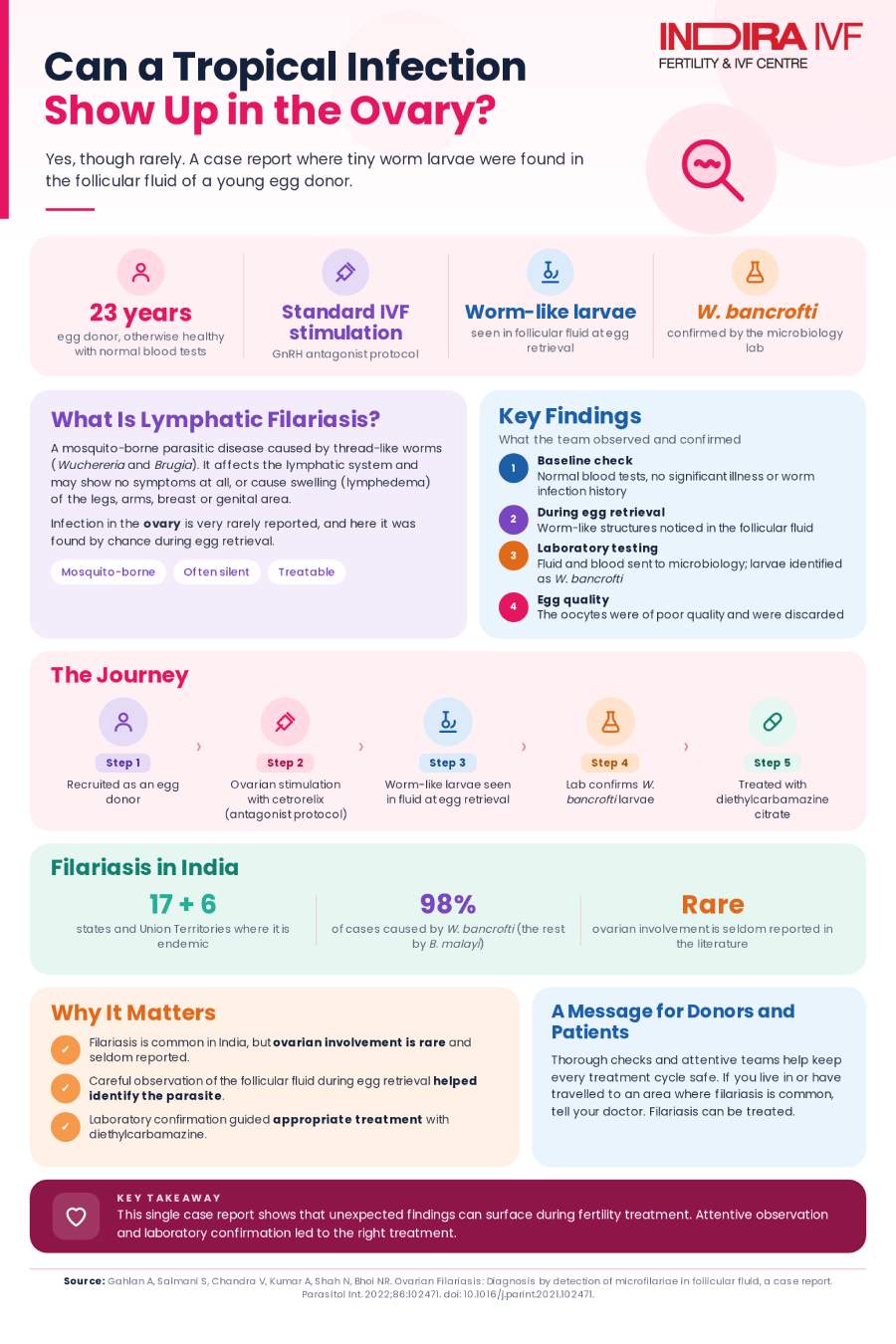
A published case report from Indira IVF documents the discovery of Wuchereria bancrofti, the parasite that causes lymphatic filariasis, in the follicular fluid of a 23-year-old egg donor during a routine IVF egg retrieval. The finding, an exceedingly rare one, explained why her eggs were of poor quality and led to prompt antiparasitic treatment.
Based on: Gahlan A, Salmani S, Chandra V, Kumar A, Shah N, Bhoi NR. "Ovarian Filariasis: Diagnosis by Detection of Microfilariae in Follicular Fluid, A Case Report." Parasitology International, 2022;86:102471. Case managed at Indira IVF Hospital, India.
Table of Contents
- Study at a Glance
- What is lymphatic filariasis, and how could it end up in the ovary?
- Key facts at a glance
- The case in detail: how the parasite was found
- Confirming the diagnosis and identifying the parasite
- Treatment and outcome for the patient
- Why ovarian filariasis is exceptionally rare
- Limitations worth knowing
- Key Results at a Glance
- What this means for patients and fertility clinics
Study at a Glance
Item | Details |
|---|---|
Evidence type | Published case report |
Patient | 23-year-old egg donor, one living child, rural background |
Clinical setting | Routine oocyte retrieval using the GnRH antagonist protocol |
Key finding | Microfilariae of Wuchereria bancrofti identified in follicular fluid |
Confirmation | Follicular fluid and blood sample analysis by the microbiology department |
Treatment | Diethylcarbamazine citrate, the standard antifilarial medication |
Main outcome | Oocytes were of poor quality and discarded; parasitic infection treated |
Publication | Parasitology International, 2022 |
DOI | 10.1016/j.parint.2021.102471 |
Lymphatic filariasis is a mosquito-borne parasitic infection that is common across India, most often producing swelling in the legs or genital area. It is rarely thought of as a fertility issue, and it is almost never discussed in the context of IVF. This case report describes an unusual and previously seldom-documented situation: a young egg donor undergoing a routine oocyte retrieval procedure whose follicular fluid, the fluid drawn from the ovary along with the eggs, was found to contain live parasite larvae. The report walks through how this was discovered, how the parasite species was confirmed, and what it meant for the donor's fertility treatment and health.
23 years old the patient, an egg donor with one living child | W. bancrofti the parasite species identified in her follicular fluid | 553 million people estimated to be at risk of lymphatic filariasis in India | Oocytes discarded due to poor quality once the infection was found |
What is lymphatic filariasis, and how could it end up in the ovary?
Lymphatic filariasis is a parasitic disease caused by thread-like worms, most commonly Wuchereria bancrofti and, less often, Brugia malayi. It spreads through mosquito bites, and adult worms typically settle in the body's lymphatic system, the network of vessels that helps manage fluid balance and immune defense. In India, the disease is considered endemic in 17 states and six union territories, with an estimated 553 million people at risk. Wuchereria bancrofti accounts for about 98% of cases in the country, with Brugia malayi responsible for the remaining 2%.
The infection can be entirely without symptoms, discovered only incidentally, or it can cause visible swelling, most classically in the legs, but also in the breast, arms, and genital area. Reports of the parasite affecting the scrotum in men are relatively common in the medical literature, but reports of it affecting the ovary, as in this case, are rare. A small number of prior case reports have described parasite larvae found unexpectedly in follicular fluid during fertility procedures, making this an uncommon but recognized, if seldom discussed, finding.
Key facts at a glance
A summary of the case described in the report:
- The patient was a 23-year-old woman from a rural area with one living child, recruited as an oocyte (egg) donor for a fertility treatment cycle.
- Her baseline blood investigations were within normal limits, and she had no history of any significant medical or surgical illness, including no known history of worm infestations.
- She underwent controlled ovarian stimulation using the GnRH antagonist protocol, a standard approach used to prevent premature ovulation during IVF and ICSI treatment.
- During the ovum pickup procedure (the process of retrieving eggs from the ovaries), her follicular fluid was found to contain worm-like structures suggestive of parasite larvae.
- The follicular fluid, along with a blood sample, was sent to the microbiology department, which confirmed the larvae to be Wuchereria bancrofti, the parasite responsible for most lymphatic filariasis cases in India.
- The oocytes retrieved were of poor quality and were discarded, and the patient was treated with Diethylcarbamazine citrate, the standard medication used to treat filarial infection.
The case in detail: how the parasite was found
The patient in this case was a 23-year-old woman with one living child, from a rural area and a low socio-economic background, who was recruited as an oocyte donor. She was of average build, and her baseline blood investigations were within the normal range, with no history of any significant medical or surgical illness. As part of a routine assisted reproduction procedure, she underwent controlled ovarian stimulation using the antagonist protocol, in which a GnRH antagonist medication (cetrorelix) is used to suppress the body's natural hormone surge and prevent early ovulation before the eggs can be retrieved.
During the ovum pickup procedure itself, the follicular fluid drawn from her ovaries revealed worm-like structures under observation, a finding that was not anticipated given her unremarkable medical history and normal baseline investigations.
Confirming the diagnosis and identifying the parasite
Once the worm-like structures were noticed, the follicular fluid was sent to the microbiology department for formal identification, along with a blood sample from the patient to help confirm the parasite species. Testing confirmed the larvae to be Wuchereria bancrofti, the species responsible for the large majority of lymphatic filariasis cases in India. This laboratory confirmation was an important step, since visual identification of worm-like structures alone would not have been sufficient to confirm the specific parasite involved or to guide appropriate treatment.
Table 1. Lymphatic filariasis in India: scale of the disease, as cited in this report
Measure | Figure |
|---|---|
States and union territories where the disease is endemic | 17 states and 6 union territories |
Population estimated to be at risk nationally | 553 million people |
Share of cases caused by Wuchereria bancrofti | Approximately 98% |
Share of cases caused by Brugia malayi | Approximately 2% |
Estimated cases of microfilariae infection nationally | Up to 27.09 million |
Estimated cases with some symptomatic presentation | Approximately 20.83 million |
Treatment and outcome for the patient
Because the oocytes retrieved during this cycle were of poor quality, they were discarded and were not used for fertility treatment. The patient was treated with Diethylcarbamazine citrate, the standard antifilarial medication used to treat Wuchereria bancrofti infection. The report does not describe any further reproductive procedures being carried out for this donor cycle beyond the discarding of the affected oocytes and the initiation of appropriate antiparasitic treatment.
Table 2. Case summary: from stimulation to treatment
Step | Finding or action |
|---|---|
Baseline evaluation | Normal blood investigations; no history of worm infestation or significant illness |
Ovarian stimulation | GnRH antagonist protocol, a standard IVF/ICSI approach |
Ovum pickup | Worm-like structures observed in follicular fluid |
Laboratory confirmation | Follicular fluid and blood sample tested; confirmed as Wuchereria bancrofti larvae |
Oocyte outcome | Poor quality; oocytes discarded |
Treatment given | Diethylcarbamazine citrate |
Why ovarian filariasis is exceptionally rare
Lymphatic filariasis most commonly affects the lymphatic system and, in men, the scrotum, where cases are reported with some regularity in the medical literature. The authors note that reports of the parasite affecting the ovary are, by contrast, rare, with only a handful of prior case reports describing similar findings of parasite larvae unexpectedly discovered in follicular fluid during fertility procedures elsewhere in the world. Filariasis is endemic across India and sub-Saharan Africa, and the authors note that its implications extend globally given how widely people travel, meaning cases can, in principle, present far from regions where the infection is typically expected.
Why this case is notable: The parasite was discovered purely as an incidental finding during a routine fertility procedure in a patient with no symptoms and no history suggestive of any worm infestation. This underscores that lymphatic filariasis can be entirely silent until it is uncovered by an unrelated medical procedure, in this instance one connected to fertility treatment rather than to any filariasis-related complaint.
Limitations worth knowing
Several factors are worth keeping in mind when interpreting this case report:
- This is a single case report describing one patient, so it cannot establish how often this kind of finding occurs among egg donors or IVF patients more broadly, particularly in regions where filariasis is endemic.
- The published report is brief, and detailed information on longer-term follow-up of the patient after antiparasitic treatment is not extensively described.
- Because the patient had no symptoms and a normal baseline evaluation, this case suggests that routine pre-treatment screening would not necessarily have identified this infection in advance, which limits how preventable this specific scenario may have been.
- As an incidental finding during an unrelated fertility procedure, this case cannot speak to how the infection might affect fertility or egg quality in other patients or under other circumstances.
Key Results at a Glance
Finding or Step | Result |
|---|---|
Worm-like structures in follicular fluid | Observed during routine ovum pickup |
Parasite species confirmed | Wuchereria bancrofti (accounts for about 98% of Indian filariasis cases) |
Oocyte quality | Poor; oocytes discarded, not used for fertility treatment |
Treatment given | Diethylcarbamazine citrate, the standard antifilarial medication |
Rarity of this presentation | Only a small number of prior case reports worldwide describe parasite larvae in follicular fluid |
These figures are drawn from a single published case report, not a comparative study, so they describe one patient's presentation rather than an expected rate of occurrence.
The reassuring takeaway: this infection, though unexpected in the setting of fertility treatment, was identified promptly through routine laboratory evaluation and treated with a standard, effective antiparasitic medication.
What this means for patients and fertility clinics
For fertility clinics, this case is a reminder that unusual findings during oocyte retrieval, even in patients with no relevant symptoms or history, are worth careful laboratory evaluation, since they can occasionally reflect a treatable underlying condition rather than a technical or procedural issue. For patients, particularly those from regions where lymphatic filariasis is endemic, this case illustrates that the infection can be entirely silent and can, in rare instances, be discovered incidentally during a fertility procedure rather than through any symptom the patient would have noticed on their own. The authors' broader message is that ovarian filariasis, while rare enough that most clinicians will never encounter it, is a recognized, if seldom discussed, cause of unexpected findings during assisted reproduction, and should be considered when unusual structures are observed in follicular fluid.
Reference: Ovarian Filariasis: Diagnosis by detection of microfilariae in follicular fluid, a case report
You May Also Want to Read
Despite A Rare Hormone Disorder and One Failed Cycle, They Finally Had a Baby Girl at 37 Weeks