
Overview
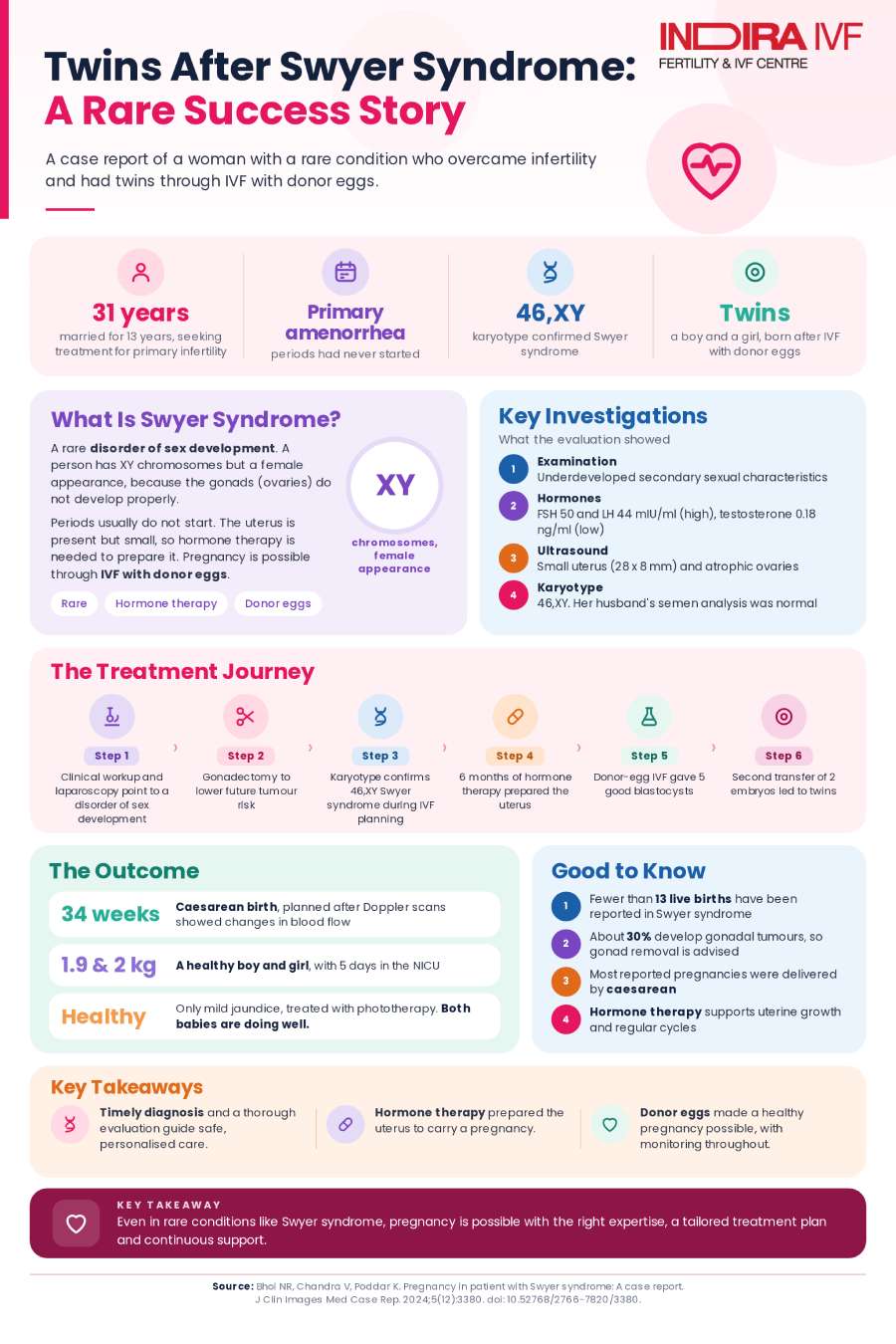
A published case report from Indira IVF documents a 31-year-old woman who had lived her whole life with Swyer syndrome, a rare disorder in which a person with male (XY) chromosomes develops as outwardly female but without functioning ovaries, undiagnosed until she sought infertility treatment. After gonad removal to reduce cancer risk, hormone therapy to prepare her uterus, and IVF using donor eggs, she carried twins to a healthy delivery.
Based on: Bhoi NR, Chandra V, Poddar K. "Pregnancy in Patient with Swyer Syndrome: A Case Report." Journal of Clinical Images and Medical Case Reports, 2024;5(12):3380. Case managed at Indira IVF Hospital, India.
Table of Contents
- Study at a Glance
- What is Swyer syndrome, and why does it complicate pregnancy?
- Key facts at a glance
- How the diagnosis was finally made
- Surgery to remove the gonads and reduce cancer risk
- Preparing the uterus for pregnancy with hormone therapy
- IVF with donor eggs and the twin pregnancy
- Limitations worth knowing
- Key Results at a Glance
- What this means for patients and families
Study at a Glance
Item | Details |
|---|---|
Evidence type | Published case report |
Patient | 31-year-old woman with primary amenorrhea and 13 years of infertility |
Diagnosis | Swyer syndrome (46,XY gonadal dysgenesis), with mixed ovarian and testicular gonadal tissue found on histopathology |
Clinical setting | IVF using donor oocytes, following bilateral gonadectomy and hormone therapy |
Treatment | Bilateral gonadectomy, hormone replacement therapy for uterine priming, double embryo transfer with donor eggs |
Main outcome | Twin pregnancy delivered by cesarean section at 34 weeks; both babies healthy |
Publication | Journal of Clinical Images and Medical Case Reports, 2024 |
DOI | 10.52768/2766-7820/3380 |
Swyer syndrome is a rare disorder of sex development in which a person has a typically male (46, XY) chromosome pattern but develops with a female outward appearance, along with gonads that fail to develop properly and do not produce eggs or the hormones needed to trigger puberty on their own. It is usually diagnosed in the late teens or early twenties, when a young woman is evaluated for absent periods. This case report describes a woman whose diagnosis was delayed until age 31, made only when she and her husband sought help after 13 years of infertility. It documents her full diagnostic journey, the surgery recommended to reduce her cancer risk, and how she ultimately carried a twin pregnancy using donor eggs.
31 years old at diagnosis, after 13 years of infertility | Fewer than 13 live births previously reported worldwide in Swyer syndrome | 5 blastocysts formed through donor-egg IVF | Twins delivered by cesarean section at 34 weeks, both healthy |
What is Swyer syndrome, and why does it complicate pregnancy?
Swyer syndrome, also known as 46, XY complete gonadal dysgenesis, is a rare disorder of sex development. People with the condition have a male (46, XY) chromosome pattern, but their gonads do not develop into functioning testes, and as a result, the body follows a female developmental path on the outside, typically with a vagina, cervix, fallopian tubes, and a uterus, alongside primary amenorrhea (periods that never start) and underdeveloped secondary sexual characteristics. Diagnosis most often occurs between ages 18 and 23, when a young woman is evaluated for the absence of periods.
Because the gonads in Swyer syndrome are dysgenetic, meaning improperly formed rather than absent, they carry an elevated risk of developing tumors, most commonly a type called gonadoblastoma, which can occur at any age, including in childhood. For this reason, gonadectomy (surgical removal of the gonads) is strongly recommended once the diagnosis is made. Removing the gonads, however, eliminates any possibility of using the patient's own eggs, meaning pregnancy can only be achieved through donor oocytes together with hormone therapy to prepare the uterus, which is the path this case report follows.
Key facts at a glance
A summary of the clinical course described in the report:
- The patient was a 31-year-old woman with primary amenorrhea, evaluated alongside her 44-year-old husband after 13 years of infertility; her husband's tests, including semen analysis, were normal.
- On examination, she had underdeveloped secondary sexual characteristics and clitoromegaly (an enlarged clitoris), with a deep vaginal canal and a palpable cervix.
- Blood tests showed elevated FSH (50 mIU/ml) and LH (44 mIU/ml), alongside a notably low testosterone level of 0.18 ng/ml, with normal thyroid and prolactin levels.
- Ultrasound revealed a hypoplastic (underdeveloped) uterus measuring 28 mm by 8 mm, with a thin endometrial lining, and both ovaries appeared atrophic.
- Diagnostic laparoscopy found a streak ovary on the left side and a gonad with testicular characteristics on the right side, located in the inguinal canal; both gonads were surgically removed due to malignancy risk.
- Karyotype analysis, performed later while planning the IVF cycle, confirmed the 46, XY chromosome pattern.
- Following six months of hormone therapy to prepare her uterus, the patient underwent IVF using donor eggs, which resulted in a twin pregnancy delivered by cesarean section at 34 weeks, with both babies healthy.
How the diagnosis was finally made
The couple, comprising a 44-year-old husband and a 31-year-old wife, sought help at an infertility clinic after 13 years of marriage. The husband's evaluation, including blood tests and semen analysis, returned entirely within the normal range. The wife's gynecological evaluation, however, revealed underdeveloped secondary sexual characteristics and clitoromegaly, while a vaginal examination found a deep vaginal canal with a palpable cervix.
Laboratory testing showed normal thyroid function and prolactin levels, but her FSH and LH, the two hormones that signal the ovaries, were both elevated, a pattern consistent with ovaries that are not responding normally. Her testosterone level was notably low. A buccal smear, a simple cheek-swab test used to check chromosomal patterns, prompted further genetic testing. Ultrasound imaging showed a hypoplastic uterus with a thin endometrial lining and both ovaries appearing atrophic, while an upper abdominal scan was otherwise normal.
Table 1. Diagnostic and genetic findings
Test | Result | What it indicates |
|---|---|---|
FSH (follicle-stimulating hormone) | 50 mIU/ml | Elevated, suggesting the ovaries are not responding normally to hormonal signals |
LH (luteinizing hormone) | 44 mIU/ml | Elevated, consistent with the same pattern of poor ovarian response |
Testosterone | 0.18 ng/ml | Notably low despite a 46,XY chromosome pattern, reflecting non-functioning gonads |
Thyroid function and prolactin | Normal | Rules out thyroid or prolactin-related causes of absent periods |
Ultrasound of the uterus | Hypoplastic uterus, 28 mm by 8 mm, thin endometrial lining | Underdeveloped uterus consistent with lifelong absence of ovarian hormone exposure |
Karyotype analysis | 46,XY | Confirms a male chromosome pattern despite a female outward appearance, establishing the diagnosis of Swyer syndrome |
Surgery to remove the gonads and reduce cancer risk
Diagnostic laparoscopy revealed a rudimentary uterus alongside bilateral normal fallopian tubes. The left gonad appeared as a streak ovary, underdeveloped tissue without functional egg-producing capacity, while the right gonad was located in the inguinal canal and showed characteristics of testicular tissue. Because of the recognized risk that dysgenetic gonads can develop tumors, bilateral gonadectomy (removal of both gonads) was performed along with salpingectomy (removal of the fallopian tubes). At the same time, the surgical team also performed excision of the phallus (an enlarged, penis-like structure) and clitoral reconstruction.
Histopathological examination of the removed tissue found ovarian stroma in the left gonad and testicular elements in the right gonad, a combination that meets the definition of true hermaphroditism, a rare condition in which both ovarian and testicular tissue are present in the same individual. This finding, together with the confirmed 46,XY karyotype, places this specific case within the broader spectrum of disorders of sex development rather than representing the most classic, textbook form of Swyer syndrome, in which both gonads are typically non-functioning streak tissue.
Table 2. Laparoscopy and gonad findings
Gonad | Location and appearance | Histopathology result |
|---|---|---|
Left gonad | Streak ovary | Ovarian stroma |
Right gonad | Located in the inguinal canal, with testicular characteristics | Testicular elements |
Preparing the uterus for pregnancy with hormone therapy
With both gonads removed, the patient's uterus needed to be prepared for pregnancy using hormone replacement therapy (HRT), since her body could no longer produce the hormones needed to support a pregnancy on its own. She received sequential exposure to estradiol valerate for 25 days each month, with dydrogesterone added during the final 10 days of each cycle. She responded well, developing regular cycles, and this HRT regimen was continued for six months before proceeding further.
A repeat ultrasound after six months showed clear growth of the uterus, to a size of 6 cm by 4 cm by 3 cm, with an endometrial thickness of 7.5 cm as reported in the study. A mock embryo transfer, a rehearsal procedure without an actual embryo, confirmed that the vaginal canal and endocervical canal were both patent (open), though the cervix was noted to be positioned high and flush with the vagina, a detail that would later factor into the pregnancy's management.
IVF with donor eggs and the twin pregnancy
Through an egg donation programme, five good-quality blastocysts were formed. The patient was first planned for an elective single embryo transfer (SET), which resulted in a chemical pregnancy loss, an early pregnancy that does not progress. Three months later, she underwent a double embryo transfer using two embryos graded 4AA and 5AB (a standard blastocyst grading system), which resulted in a twin pregnancy.
Because of concerns about uterine muscle strength given her high-positioned cervix, an abdominal cerclage, a procedure to reinforce and support the cervix during pregnancy, was planned and performed, which she tolerated well. The antenatal period was otherwise uneventful. At 34 weeks, Doppler ultrasound changes suggested compromised blood flow, prompting a planned cesarean section (LSCS). One healthy male baby and one female baby were delivered, weighing 1.9 kg and 2 kg respectively. Both were kept in the neonatal intensive care unit (NICU) for five days before being moved to their mother's side. The postoperative period was uneventful, with no major neonatal complications apart from physiological jaundice, which resolved with phototherapy. Both babies are reported to be doing well.
Limitations worth knowing
Several factors are worth keeping in mind when interpreting this case report:
- This is a single case report describing one patient, so it cannot establish how often successful pregnancies occur in women with Swyer syndrome or similar disorders of sex development, or predict outcomes for any individual patient.
- The histopathology in this case found both ovarian and testicular tissue across the two gonads, meeting the definition of true hermaphroditism. This means the case sits within a broader spectrum of disorders of sex development, and the findings may not generalize to more classic presentations of Swyer syndrome with purely streak gonads.
- The authors note that fewer than 13 live births have been documented worldwide in women with Swyer syndrome, meaning the overall evidence base for pregnancy outcomes in this population remains very limited.
- Long-term follow-up of the mother's health after pregnancy, including ongoing cancer surveillance and bone health on hormone therapy, is not detailed beyond the immediate postoperative and postpartum period described in this report.
Key Results at a Glance
Measure | Result |
|---|---|
Hormone levels at diagnosis | FSH 50 mIU/ml, LH 44 mIU/ml, testosterone 0.18 ng/ml |
Uterus size before hormone therapy | 28 mm by 8 mm, thin endometrial lining |
Uterus size after 6 months of hormone therapy | 6 cm by 4 cm by 3 cm, with an endometrial thickness of 7.5 cm as reported |
Karyotype | 46,XY confirmed (performed later, while planning the IVF cycle) |
First embryo transfer (elective SET) | Chemical pregnancy loss |
Second embryo transfer (double ET, donor eggs) | Twin pregnancy achieved |
Delivery | Cesarean section at 34 weeks; babies weighing 1.9 kg and 2 kg, both healthy after a 5-day NICU stay |
These figures are drawn from a single published case report, not a comparative study, so they describe one patient's course of treatment rather than an expected outcome for others with this condition.
The reassuring takeaway: despite a male chromosome pattern and non-functioning gonads, this patient's uterus responded normally to hormone therapy and successfully carried a twin pregnancy to a healthy delivery, using donor eggs.
What this means for patients and families
For women diagnosed with Swyer syndrome or a similar disorder of sex development, this case offers a considered, real-world illustration of what fertility treatment can look like. The authors emphasize that a 46,XY chromosome pattern does not prevent the uterus from responding normally to hormone therapy or from supporting a pregnancy, meaning that donor-egg IVF, combined with appropriately timed hormone replacement therapy, offers a realistic path to pregnancy once functioning ovaries are absent. At the same time, gonadectomy remains an important recommendation following diagnosis, given the elevated risk of gonadal tumors, even though it removes the possibility of using one's own eggs. The authors also note that cesarean delivery is more likely in this population, often due to a hypoplastic uterus and pelvic structural factors, as was the case here. Because this diagnosis can be delayed into adulthood, as it was in this patient until age 31, thorough evaluation for anyone presenting with primary amenorrhea and infertility, regardless of age, is an important takeaway for clinicians and patients alike.
Reference: Pregnancy in patient with Swyer syndrome: A case report.
You May Also Want to Read