
Overview
A published case report from Indira IVF follows a pregnancy in which a first-trimester scan showed a nuchal translucency measurement well above the typical threshold for concern. After thorough genetic testing and continued monitoring found no underlying abnormality, the pregnancy proceeded to a healthy delivery, and the child remains developmentally normal four years later.
Based on: Bhoi NR, Chandra V, Murdia K, Jhamb T. "Isolated Increased Nuchal Translucency With Normal Chromosomal Study: A Case Report." Cureus, 2023;15(11):e49209. Case managed at Indira IVF Hospital, India.
Table of Contents
- Introduction
- What is nuchal translucency, and why does an elevated reading raise concern?
- Key facts at a glance
- The prenatal findings in this case
- How genetic testing ruled out chromosomal causes
- Continued monitoring through pregnancy
- Delivery and long-term outcome
- What broader research says about isolated increased NT
- What this means for expectant parents
Introduction
Nuchal translucency (NT) is a measurement taken during a first-trimester ultrasound of the small pocket of fluid normally present behind a developing fetus's neck. Because an unusually thick NT measurement is statistically associated with chromosomal conditions and structural abnormalities, it has become one of the most widely used early screening markers in prenatal care. This case report follows a pregnancy conceived through IVF in which the NT measurement was markedly elevated, a finding that can understandably alarm expectant parents and raise the question of whether to continue the pregnancy. The report documents, step by step, how a thorough evaluation distinguished a screening flag from an actual diagnosis, and it argues for measured, well-informed counseling before any decision about the pregnancy is made.
Study at a Glance | ||||
Item | Details | |||
Evidence type | Published case report | |||
Patient | 33-year-old woman, pregnancy conceived through IVF and ICSI with single embryo transfer | |||
Key finding | Nuchal translucency of 5.6 mm at 12 weeks, well above the typical concern threshold | |||
Clinical setting | Prenatal genetic testing and monitoring following an elevated first-trimester scan finding | |||
Evaluation | Amniocentesis with FISH and full karyotype, serial ultrasounds, fetal echocardiography, postnatal microarray | |||
Main outcome | Healthy term delivery; no chromosomal abnormality; normal development at 4 years | |||
Publication | Cureus, 2023 | |||
DOI | 10.7759/cureus.49209 | |||
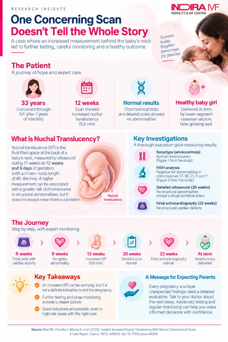
5.6 mm nuchal translucency at 12 weeks (typical concern threshold: about 3 mm) | 46 normal chromosomes confirmed by genetic testing | Term delivery healthy baby girl, via cesarean section | 4 years followed up with no developmental abnormality | |
What is nuchal translucency, and why does an elevated reading raise concern?
Nuchal translucency refers to a normal, fluid-filled space that appears behind a fetus's neck on ultrasound during a specific window of the first trimester, typically between 11 weeks and 12 weeks and six days of pregnancy, when the fetus measures between 45 mm and 84 mm from crown to rump. Every fetus has some degree of NT, but when the measurement is unusually thick, it becomes a statistical marker associated with a higher likelihood of chromosomal abnormalities (such as Down syndrome), structural birth defects, and, in more severe cases, pregnancy loss.
Published research cited in this case report indicates that fetal abnormalities associated with increased NT can range from 1.6% to 45%, and when the measurement exceeds the 99th percentile for gestational age, the prevalence of genetic disorders may range from 0.5% to 6.6%. One cited study found that fetuses with increased NT experienced an adverse pregnancy outcome, including miscarriage, intrauterine death, or termination, in 17.8% of cases, compared with just 1.5% among fetuses with a normal measurement. These figures explain why an elevated NT reading is taken seriously, but as this case illustrates, they also leave considerable room for a favorable outcome once other conditions have been ruled out.
Key facts at a glance
A summary of the clinical course described in the report:
- The patient, a 33-year-old woman, conceived through IVF and ICSI with single embryo transfer after seven years of primary infertility, with no family history of genetic disorders on either side.
- At 12 weeks of gestation, ultrasound showed a nuchal translucency measurement of 5.6 mm, well above the typical threshold of concern (commonly cited as 3.0 to 3.5 mm), with the fluid extending down toward the lower back area.
- Despite the high NT, several reassuring signs were present at the same scan: the nasal bone was visible, there was no tricuspid valve regurgitation, and blood flow through the ductus venosus (a key fetal blood vessel) was normal.
- Genetic testing on an amniocentesis sample, examining chromosomes 13, 18, 21, X, and Y across 200 cells, found no evidence of any chromosomal deletion or duplication, with a completely normal set of 46 chromosomes.
- A later scan at 16 weeks raised the possibility of a cystic hygroma (a related fluid-filled swelling), but the parents, after counseling, chose to continue the pregnancy.
- Fetal echocardiography at 22 weeks found no congenital heart defect, and a detailed anatomy scan found no structural abnormality apart from a single umbilical artery (a variant that is not uncommon and often has no clinical significance on its own).
- The baby girl was delivered at term by cesarean section, and a postnatal genetic test (microarray) confirmed no chromosomal abnormality. She has since completed four years of life with no developmental, physical, or psychological abnormality.
The prenatal findings in this case
The pregnancy's first trimester began without concern: an ultrasound at six weeks confirmed a fetal pole with cardiac activity and a yolk sac, and a follow-up scan at nine weeks showed no obvious fetal abnormality. The pivotal finding came at the 12-week scan, when the NT measurement was recorded at 5.6 mm, with the associated fluid extending beyond the neck toward the lower back, a more extensive presentation than an isolated neck swelling. At the same visit, however, several other markers examined during this type of scan, including the nasal bone, tricuspid valve, and ductus venosus blood flow, were all found to be normal, which are generally regarded as favorable signs even when the NT itself is high.
Table 1. Timeline of key prenatal findings
Gestational stage | Finding |
|---|---|
6 weeks | Fetal pole with cardiac activity and yolk sac seen; no concerns |
9 weeks | No gross fetal abnormality on ultrasound |
12 weeks | Nuchal translucency 5.6 mm; nasal bone visible; no tricuspid regurgitation; normal ductus venosus flow; single umbilical artery noted |
16 weeks | Nuchal thickness 4.6 mm; features suggestive of a cystic hygroma; parents chose to continue the pregnancy after counseling |
20 weeks | Detailed anatomy scan showed no structural abnormality apart from the single umbilical artery |
22 weeks | Fetal echocardiography showed no congenital heart defect; brain and spine structures appeared normal |
How genetic testing ruled out chromosomal causes
Because of the significantly elevated NT, the patient was advised to undergo a fetal karyotype, a test that examines the number and structure of chromosomes, obtained through amniocentesis (a procedure that samples the fluid surrounding the fetus). The specific technique used, called fluorescent in situ hybridization (FISH), specifically screened chromosomes 13, 18, 21, X, and Y, the chromosomes most commonly associated with the conditions linked to increased NT, such as Down syndrome (chromosome 21) and Edwards syndrome (chromosome 18).
Table 2. Genetic testing results
Test | What it checked | Result |
|---|---|---|
FISH analysis (amniocentesis) | Chromosomes 13, 18, 21, X, and Y across 200 cells | 100% of cells negative for deletion or duplication |
Full karyotype (GTG banding) | All 46 chromosomes, at a resolution of 550 | Normal karyotype; no sex chromosome abnormality detected |
Postnatal microarray (blood sample) | Broader screen for chromosomal abnormalities after birth | No chromosomal abnormality found |
Every test performed, both before and after birth, returned a normal result. This consistent picture across multiple testing methods, rather than a single reassuring test, is what allowed the care team to progress with more confidence through the rest of the pregnancy.
Continued monitoring through pregnancy
Rather than relying on the genetic test result alone, the pregnancy was followed with a series of additional, targeted evaluations. At 16 weeks, the persistence of fluid accumulation raised the possibility of a cystic hygroma, a soft, spongy swelling most often found on the neck but which can also appear elsewhere on the body. This finding can be associated with a range of underlying conditions, so it warranted careful counseling of the parents, who, after being informed of the normal genetic results and the plan for continued monitoring, chose to proceed with the pregnancy.
The subsequent 20-week detailed anatomy scan found no structural abnormality beyond the single umbilical artery, and the 22-week fetal echocardiography, a specialized ultrasound of the fetal heart, ruled out any congenital heart defect. This scan also assessed brain and spinal structures in detail, all of which appeared normal. Taken together, this layered approach, combining genetic testing with repeated, targeted imaging, provided a fuller picture than any single test could have offered on its own.
Delivery and long-term outcome
The remainder of the pregnancy was uneventful. The patient delivered a term female baby by lower-segment cesarean section. Following delivery, a microarray test on a blood sample from the baby, a more sensitive genetic screen than the prenatal testing performed, again found no chromosomal abnormality. At the time of this report's writing, the child had completed four years of life without any developmental, physical, or psychological abnormality.
What broader research says about isolated increased NT
This case report situates its findings within a wider body of research on increased NT with a normal karyotype. One cited study found that among fetuses with an NT of 3.5 mm or greater and a normal karyotype, the overall risk of an adverse pregnancy outcome was 25.9%, meaning the overall chance of a favorable outcome was 74.1%. Another line of research has suggested that the threshold used to recommend invasive testing might reasonably be lowered from 3.5 mm to 3.0 mm, reflecting ongoing refinement of these risk estimates.
An important caveat the report raises: even with today's standard prenatal diagnostic testing, more than 80% of cases of increased NT do not yield a specific causal finding. Separately, research cited in the report found that pathogenic copy number variations, small genetic changes not always detected by standard karyotype testing, were found in only 0.8% to 5.3% of fetuses with isolated elevated NT, and it is these that would account for some of the less favorable outcomes seen even when initial testing appears normal.
One study referenced in the report followed 55 cases of increased NT with no aneuploidy (78% of the group studied) and found a favorable pregnancy outcome at term in 40 cases, representing 56% of the total group and 72% of the pregnancies with normal chromosomes. This body of evidence, taken as a whole, supports the central message of the present case report: a high NT reading raises the statistical likelihood of complications, but with normal genetic testing and careful follow-up, most such pregnancies can still progress to a healthy outcome.
What this means for expectant parents
For parents who receive an elevated NT result, this case offers a considered, real-world illustration of how such a finding can play out. The authors conclude that NT assessment should be a routine part of first-trimester prenatal care, and when the measurement is elevated, the appropriate response is increased surveillance and specialized ultrasound screening, not an automatic assumption of a poor outcome. The report specifically emphasizes that proper counseling before the first scan, transparent discussion of all test results as they become available, and compassionate, reassuring guidance throughout the evaluation process are essential to avoiding the termination of a pregnancy that may, as in this case, prove entirely healthy. Every case is different, and this report should be read as one illustrative account rather than a guarantee of outcome, underscoring why thorough individualized evaluation matters more than the initial measurement alone.
Key Results at a Glance
Finding or Test | Result | Interpretation |
|---|---|---|
Nuchal translucency (12 weeks) | 5.6 mm | Well above the typical threshold of concern (about 3.0 to 3.5 mm) |
FISH analysis (amniocentesis) | Normal in 100% of 200 cells screened | No deletion or duplication in chromosomes 13, 18, 21, X, or Y |
Full karyotype | Normal, 46 chromosomes | No sex chromosome abnormality detected |
Fetal echocardiography (22 weeks) | Normal | No congenital heart defect |
Postnatal microarray | Normal | Confirms no chromosomal abnormality after birth |
Developmental follow-up | 4 years, no abnormality | No developmental, physical, or psychological issues identified |
These figures are drawn from a single published case report, not a comparative study, so they describe one pregnancy's course rather than an average expected outcome.
The reassuring takeaway: an elevated nuchal translucency reading is a screening flag, not a diagnosis. In this case, consistent normal results across multiple genetic and structural tests, over time, supported continuing the pregnancy, which proceeded to a healthy outcome.
Reference: Isolated Increased Nuchal Translucency With Normal Chromosomal Study: A Case Report
More Fertility Research to Explore