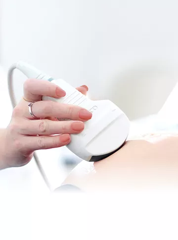
Ultrasound is a indirect vision for fertility testing. Ultrasound is now an essential part of infertility testing and treatment.
Ultrasound is a indirect vision for fertility testing. Ultrasound is now an essential part of infertility testing and treatment. Ultrasound was invented by Obstetrician Ian Donald and Engineer Tom Brown in 1950. Ultrasound was used in clinical practice in Glassgow Hospital since 1956. Ultrasound scan works by using high frequency sound waves to create an image of internal organs.
Majority of the ultrasound are done transvaginal i.e. via vagina with specialized probe in fertility testing and treatment; as it gives better visualization and magnification of pelvic organs for better diagnosis and management.
A. Uterus:-
1. Assess endometrial cavity where actual pregnancy grows i.e. endometrial size , shape and thickness
2. Rule out Adenomyosis and Fibroid which are responsible for infertility and pregnancy losses.
3. 3D imaging of uterine shape and volume, rule out uterine developmental defects.
4. Rule out Endometrial polyps and uterine septum {partial/complete} If found on ultrasound we can plan hysteroscopic removal prior embryo transfer.
B. Ovaries:-
1. Evaluate ovarian reserve {anterior follicle count } so that further mode of management {IUI/IVF-OPU/OD}can be decided and discussed with couple after correlating with AMH level.
2. Rule out type and size of cyst { simple/ haemorrhagic/chocolate/endometrioma} also rule out pelvic pathology like T-O Mass.
C. Fallopian Tubes:-
Normal healthy fallopian tubes cannot be visualized on ultrasound.
1. Partial or complete Hydrosalphinx can be diagnosed if it is swollen or filled with fluid.
2. Sono hysterogram { or Saline Infusion Sonography } is done using ultrasound to check – shape of the uterus, rule out uterine abnormality and adhesions, check patency of fallopian tubes .
D. Vaginal Doppler Study:-
Ultrasonographic parameters and Doppler analysis of uterine bloodflow can be of value in the prediction of endometrial receptivity in infertile female patients undergoing embryo transfer .Uterine scoring system included all the following parameters :Endometrial morphology,Endometrial blood flow within zone 3,Myometrial echogenicity,Uterine Artery Pulsatility Index,End diastolic blood flow and Myometrial blood flow internal to arcuate vessels.
Pregnancy rates were higher in women with thick distinct five line endometrium and multifocal endometrial vascularity within zone 3.Absent endometrial flow despite higher values for other parameters was associated with no conception.
A. Ovary :-
1. In natural cycle , to monitor follicle development and once any follicle achieves size of 18 to 22 mm , advice patient for Timed intercourse. Also we can confirm on ultrasound whether growing follicle has ruptured or not
2. In IUI cycle, follicular monitoring is from day 2 of periods with oral / injectable ovulation inducing drugs. Trigger is given when optimum size of any single follicle is achieved and then IUI is planned accordingly.
3. In IVF {self cycle} –
a. On day 2 of period a baseline ultrasound is performed , to examine ovaries, number of follicles are counted and their size is measured, depending on that a dose is decided ( considering age, BMI, AMH ) for hormonal stimulation.
b. Monitor response to stimulation of injection by transvaginal route frequently.
c. Once adequate size of maturation of follicles is achieved 18 to 22mm final maturation trigger is given by HCG/ Decapeptyl depending on cohort number.
To confirm cohort size we can do 3D ultrasound imaging of cohort.
d. 36 Hours after taking trigger shot, oocyte retrieval is done which involves removing eggs from ovary under anaesthesia using transvaginal ultrasound.
B. Uterus :-
1. To assess endometrial thickness in natural / IUI cycle/ IVF cycle (fit to transfer)
2. Embryo transfer is done under abdominal ultrasound guidance in IVF. A catheter tube is inserted in uterine cavity to place chosen embryo for transfer.
3. To study effect of estrogen on endometrial thickness in IVF cycle.
4. To confirm pregnancy on ultrasound after β-hcg report, rule out intra or extra uterine pregnancy, number of pregnancies (single or multiple).
Ultrasound is non invasive , easier , safe , cheap , and less time consuming modality for fertility testing and treatment . It has revolutionised the investigative approach and treatment of an infertile couple.
2023
Female Infertility Male Infertility
While it depends on the correct circumstances and the stage of the woman's men...
2023
Estrogen is a very important hormone in a female’s body. It is especially ne...
2022
Female Infertility Infertility Problems
As we all know infertility rate is constantly rising in our society day by day...
2022
Female Infertility Infertility Problems
Pregnant women and those in close-contact with them must take necessary precau...
2022
Female Infertility Infertility Problems
What do we know today about the benefits and risks of vaccination in pregnant ...
2022
Female Infertility Infertility Problems
A lot of people get themselves examined once or twice a year by their doctor. ...
2022
Female Infertility Infertility Problems
It is a common belief over decades that women with normal menstrual cycle patt...
2022
Female Infertility Infertility Problems
Fertilization- Fertilization is a complex process where a sperm and egg uni...
2022
Female Infertility Infertility Problems
Infertility is inability to conceive after one year or more with regular unprote...
2022
Infertility Problems Female Infertility
Incidence of infertility in India is 10 to 15%, in other words 27.5 million co...
Get quick understanding of your fertility cycle and accordingly make a schedule to track it