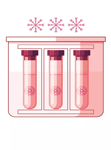
Embryo cryopreservation has diminished the number of fresh embryo transfers and maximized the effectiveness of the IVF cycle.
The very first successful mouse embryo cryopreservation (CP) was reported individually from one another by two research groups in the year 1972. Twelve months later, the birth of the first calf through frozen embryo was posted. The 1st human pregnancy from a frozen embryo was achieved with the same procedure applied successfully for CP of mouse and cow embryos; however, it had been terminated simply by spontaneous abortion in the second trimester. Since then, embryo CP,has become routine procedure in human assisted reproduction (AR) as well as oocyte CP is getting introduced into clinical training and is also getting more and more widely used. Those couples opting for cryopreservation often ask our experts can you explain some advantages of cryopreservation. What does embryo cryopreservation cost? What are embryo cryopreservation risks? Is cryopreservation of embryos in animals possible? Can you define oocyte cryopreservation? What is embryo vitrification? Describe ovarian tissue cryopreservation? What is the cryopreservation of eggs? Can you explain oocyte cryopreservation success rates? How long can a woman freeze her eggs? Is there any oocyte cryopreservation near me? What is the relation between Cryopreservation and IVF success? Although it is an abysmal subject, the best IVF specialist in Amritsar Dr. Shilpa Gulati at the Indira IVF clinic in Amritsar has tried to cover all the above queries in a concise manner in the given below treatise.
Wilmut I, Rowson LEA. Experiments on the low temperature preservation of cow embryos. Veterinary Record. 1973;92(26):686–690.
Dr. Suman Gynecologist & IVF Specialist Explained that: “Embryo cryopreservation has diminished the number of fresh embryo transfers and maximized the effectiveness of the IVF cycle.” Likewise, embryo CP is a crucial tool in cases of terminated embryo transfer (ET) due to the chance of an ovarian hyperstimulation, endometrial bleeding, elevated serum progesterone levels on the day of triggering, or any other unplanned events. There is still a sizable debate on the best stage, protocol, and cryoprotective additives (CPA) to utilize. The average prospect of a frozen stored embryo to become a living child lies in the order of 4%, and babies born from cryopreserved embryos do not represent more than 8−10% of the entire number of babies born from assisted reproduction. Nevertheless, it is unquestionable that successful CP of zygotes or embryos has dramatically increased the medical benefits and increasing conceiving rates possible for partners following a single routine of ovarian stimulation and IVF. Results presented as the addition of the delivery rate per oocyte harvested varies significantly in the literature, between 2% and 24%. The data demonstrates women who had fresh and frozen transfers of embryos obtained 8% additional births by using their cryopreserved embryos stipulated the best IVF specialist in Amritsar Doctor Shilpa Gulati at the Indira IVF and infertility treatment center in Amritsar.
Typically the metaphase II (MII) oocyte has a very unique structure (i.e., enormous size, very sensitive to low temperature, extremely delicate, high water content, lower surface to volume ratio, existence of the spindle and other cell organelles, not optimal plasma membrane layer permeability to CPA and water, etc.) which results in complex difficulties connected with its CP. The particular spindle is important regarding the events following fertilization in the completion of meiosis, second polar body formation, migration of the pronuclei, and formation of the first mitotic spindle. Typically the damage (depolymerization) and lack of the spindle compromise the ability of the specific oocyte to fertilize and undergo average preimplantation growth. Additionally, hardening of the particular zona pellucida—which is the consequence of CP—can negatively affect the normal fertilization process. However, oocyte cryopreservation offers more advantages when compared with embryo freezing:
(1) Virility preservation in women vulnerable to losing fertility due to oncological treatment, premature ovarian failure, or chronic illness;
(2) It can assist alleviate religious as well as other moral, legal, and ethical issues of embryo storage;
(3) It helps to get over problems such as whenever the husband is not able to produce a feasible sperm sample or whenever spermatozoa cannot be identified in the testis at a given moment in case there is nonobstructive azoospermia;
(4) It makes egg banks and egg donations possible by removing donor-recipient synchronization problems; plus
(5) It allows ladies to postpone childbirth right up until a later time or age (e.g., after establishing a new career, and so forth).
Typically the latter is called interpersonal freezing when the oocytes are cryopreserved for nonmedical purposes. For about ten years, in parallel along with the technical improvement of oocyte freezing, the probability of egg storing concerning nonmedical objectives is even more extensively discussed and more commonly accepted by the general population and professional committees in the U.S and Europe. The goal of the social freezing is to prevent age-related fertility decline, which is usually widely promoted by fertility clinics and the lay (un-academic) press throughout the globe. It is a new fact that the reproductive performance or ability of females is about their 25–30 years of age. Afterward, maternity rates decline relatively quickly from 35 years, plus miscarriage rates rise significantly.
After the age of 43 years, the chances of becoming pregnant are incredibly lower. It is a global tendency that ladies decide to give birth at later stages, as in contrast to 20–30 years back. Information of our patients getting frozen cycle indicate the average age (n = 3601) increased from 31.8 to 35.4 in the last ten years responded Doctor Shilpa Gulati at the Indira IVF as well as test tube baby treatment center in Amritsar.
The effect of age is noticeable within the frozen embryo survival rate which slowly yet consistently decreased in the last ten years since the average age of the particular patients increased by four years without doing any kind of modification within the freezing procedure (89% versus 81%; P < 0.0001). The number of active frozen cycles is considerably lower over 30 years, and there is a substantial significant difference over thirty-five compared with less than 30 years of age (P < 0.01 and P < 0.0002).
The achievement rate of embryo/oocyte CP depends upon is determined by several variables: efficiency of the freezing procedure, carriers used for vitrification (open versus closed), regularity of cycles with CP in the assisted reproductive system program, the conditions regarding selection of embryos or oocytes for freezing, and the outcomes of fresh embryo transfers. Results could be expressed as survival rates (but that is not enough only, retention of normal physical function of the cellular organelles is essential), implantation rates, pregnancy rates, or even delivery rates per transmitted or thawed embryos or harvested oocytes informed the fertility doctors in Amritsar.
Typically the traditional slow cooling procedures for CP are known as equilibrium cooling as well as the rapid/ultra T quick procedures (vitrification) as no equilibrium cooling. Various factors influence the survival of embryos in addition to oocytes cryopreserved by equilibrium or no equilibrium cooling methods.
The most delicate challenge during the CP of embryos and oocytes is to stop the creation of ice crystal in addition to toxic concentrations of solutes, which are the two leading causes of cellular death connected with CP, although maintaining the functionality of intracellular organelles and the particular viability of the embryo or oocyte. To carry out, therefore, the freezing solution, within which the cells are usually suspended, must be supplemented with cryoprotective additives (CPA). Exposure to CPA facilitates the dehydration of the particular cell and reduces intracellular ice formation. The CPA may be split up into a couple of groups: intracellular/membrane-permeating (i.e., propylene glycol/PG/, DMSO, glycerol/G/, and ethylene glycol/EG/) in addition to extracellular/membrane-no permeating compounds (i.e., sucrose, trehalose, glucose, amid, ficoll, proteins, and lipoproteins).
The permeable CPA displaces water via an osmotic gradient and partly occupies the place of the intracellular water, while the extracellular CPA increases the extracellular osmolarity generating an osmotic gradient across the particular cell membrane supporting the dehydration of the cell before CP. At the same time, it helps prevent the rapid entry of water into the cell after thawing during rehydration/dilution out of the permeating CPA told Dr. Shilpa Gulati at the Indira IVF fertility treatment center in Amritsar.
Dehydration of the cell mainly depends on the permeability properties of the cellular membrane. There are variations in permeability among the embryos of different species to water and permeating CPA. Embryos usually are less permeable to G to PG or EG. Furthermore, the earlier the stage of development, typically the less porous is the particular embryos. The permeability properties of premature and mature oocytes fluctuate and can vary simply by 7-fold between individual MII oocytes.
This difference within membrane permeability may have a powerful impact on the particular outcome of slow freezing of oocytes but could be controlled by the elevation of the concentration of the non-permeable CPA and the environmental temperature. By getting the level of non-permeating CPA increased (sucrose: 0.2 and 0.3 M) higher survival rates had been reported, and the general fertilization rates of frozen-thawed oocytes seemed to be similar to those of fresh oocytes stated Dr. Shilpa Gulati at the Indira IVF hospital in Amritsar.
Before slow cooling, dehydration of the embryos/oocytes is usually carried out by direct exposure to a combination of permeable and non-permeable CPA (duration: ten minutes). When it comes to human embryos/oocytes, with few exceptions, lower concentration of PG (1.5 M) and sucrose (0.1–0.25–0.5 M) is used for early cleavage stage embryos and oocytes and G for blastocyst stage embryos. In case there is the original successful CP protocol mouse, and cow embryos were cooled with a slow cooling rate (between minus 0.3°C–0.5°C/min) to shallow temperature ranges of minus 80°C–120°C. Consequently, the length of the procedure had been very long (several hours). Willadsen and Willadsen et al.described a variation of this specific method in which sheep and bovine embryos had been cooled slowly at a new rate of 0.3°C/min but just to minus 30–35°C before being plunged directly into liquid nitrogen (LN2). With this specific modification, the duration of the CP process was considerably shortened (1.0–1.5 hours).
Since then, this particular short protocol has become the treatment of choice for freezing of domestic animal embryos. Despite the outstanding results achieved with animal embryos, human embryos are often frozen with a reduced cooling rate of 0.3°C/min to about minus 30°C to 40°C, then by an elevated cooling rate of minus 50°C/min to a temperature of minus 80°C–150°C before being plunged into LN2. During slow cooling, the dehydration process is usually thought to continue right up until minus 30°C, after which often any remaining water will be super cooled. Throughout the slow cooling stage ice nucleation (seeding) will be induced manually between −5 and −8°C (close to the actual freezing point of the solution). Embryos/oocytes cooled slowly to subzero temperatures of minus 30°C to 40°C before getting super cooled to minus 196°C require rapid warming/thawing in the warm water of 25°C–37°C educated Dr. Shilpa Gulati at the Indira IVF hospital in Amritsar.
Rapid thawing is adopted by removal of the CPA from the embryo or oocyte. Rehydration of the cells is usually carried out in reducing concentrations of permeating CPA, generally in the existence of increased levels of non-permeating CPA. A typical medical practice is to dilute CPA out of the particular frozen embryo or oocyte in a stepwise fashion. The use of a non-permeating solute, such as sucrose as an osmotic barrier, decreases the chances of an osmotic shock plus shortens the duration of the particular process (see earlier). Long term storage of embryos and oocytes requires temperatures below minus 130°C, the glass changeover temperature of the water. In medical practice, the simplest and most secure way would be to store cryopreserved embryos and oocytes in LN2 at minus 196°C. Mouse model experiments indicate that the extended storage of embryos/oocytes does not affect the results of thawed cycles. Live mice plus sheep have been developed from cryopreserved embryos kept for more than fifteen years in LN2. Children have been born from embryos that were cryopreserved for over eight and 12 years assured Doctor Shilpa Gulati at the Indira IVF fertility treatment center in Amritsar.
Vitrification is an alternative method to embryo and oocyte cryopreservation which often has been recently referred to as a revolutionary technique; on the other hand, the very first successful embryo vitrification was published around typically the middle of the eighties. Vitrification is unique from slow freezing for the reason that it avoids the creation of ice crystals inside the intracellular as well as extracellular space. It is the solidification of a solution by a significant elevation in viscosity in low temperatures without ice crystal formation, a procedure attained by a blend of a high concentration of CPA (4–8 mole/L) in addition to an extremely high (ultra-rapid) cooling rate. In contrast to slow freezing (when dehydration in the embryos and oocytes starts in the course of the equilibration in the particular freezing solution just before slow cooling and continues throughout slow cooling to minus 30–35°C). During vitrification, cells are dehydrated mainly just before the start of the ultra-rapid cooling by direct exposure to large high concentrations of CPA, which is essential to obtain a vitrified intracellular and extracellular state soon after. The actual risk associated with the vitrification procedure is usually the high concentration of CPA that could end up being toxic to cells.
Nevertheless, it is possible to limit CPA toxicity by mixing different CPA, thus decreasing the relative focus of each CPA, and by merely reducing the exposure period of embryos and oocytes towards the solution to a minimum. The freezing solutions which are frequently used for vitrification are usually composed of permeating (e.g., EG, G, DMSO, PG, acetamide; > 4M) and non-permeating (e.g., sucrose, trehalose > 0.5 M) agents. In some protocols, the vitrification medium is likewise supplemented with macromolecules like polyethylene glycol, ficoll, or polyvinylpyrrolidone. By improving viscosity, the macromolecules help vitrification with lower levels of CPA. To further enhance the cooling rate (> 10.000°C/min) essential for effective vitrification, the volume of the solution in which the particular embryos and oocytes are vitrified has been recently drastically lowered (0.1–2 μL). Hence, to accomplish this, specific carrier systems (open vs. closed) have been created such as free pulled straws, flexipet-denuding pipettes, Cryotop, electron microscopy copper grids, cryoloops, or even the Hemi-Straw system.
Closed methods have been developed for safety reasons. Comparing the particular open and closed methods Bonetti et al.using closed carriers reported acceptable survival rates, but with numerous vesicles throughout the cytoplasm of oocytes which is often the likely consequence of not rapid enough temperature lowering in the closed method. Nevertheless, as a result of the bettering results, typically the application of vitrification—especially about cryopreservation of human blastocyst and oocyte—has recently been significantly increased articulated Dr. Shilpa Gulati at the Indira IVF and test tube baby treatment center in Amritsar.
Technically vitrification is challenging to perform, as a result of the concentrated, viscous, and small volume of vitrification solutions wherein the embryos and oocytes must end up being handled for only a minimum period (<1 min) ahead of and through vitrification. Therefore, to achieve the optimal/high survival rate, the embryologist carrying out vitrification must be very well trained. It is not necessarily the situation in the circumstance of slow freezing if the embryos and oocytes are cooled gradually (with a particular cell freezer), because slow freezing is a more flexible approach. Similarly to slow freezing, rapid thawing is needed concerning the optimal survival of vitrified embryos and oocytes, followed simply by stepwise rehydration using related techniques employed after slower cooling reasoned Dr. Shilpa Gulati at the Indira IVF center in Amritsar.
Generally, PG is employed for the freezing of zygote and cleavage stage embryos and G for your cryopreservation of blastocysts. For many years, the recommended stages for human embryo cryopreservation had been the zygote and early cleavage stages. Blastocyst freezing was abandoned for many years}, considering that only 25% of zygotes were able to attain the blastocyst stage in vitro in usual culture media, and overall reduced pregnancy rates were noted. In a new embryo culture systems like the co-culture on feeder cells in addition to the sequential media—have newly been developed so that it is possible to obtain high-quality blastocysts within 50–60% of the instances. Therefore, the significance of blastocyst Cryopreservation elevated within the last 8–10 years. Moreover, several published data reveal that human blastocysts attained using sequential media seem to be only 50 percent as cryoresistant as the cocultured types emphasized Doctor Shilpa Gulati at the Indira IVF clinic in Amritsar.
The early cleavage phase embryos are considered surviving cryopreservation if they keep at least 50% of their preliminary blastomeres intact after thawing. The moderate loss in cells did not considerably affect implantation. In an earlier, sizeable multicenter study along with 14000 cleavage stage slower frozen and thawed embryos it had been determined that 73% of the embryos had at least half their preliminary blastomeres still intact and also the results showed medical pregnancy and implantation rates of 16 and 8.4%, respectively, after transfer. In another study associated with over 300 single frozen embryo transfers of Day 2 embryos at the 4-cell stage and the particular embryos lost only a single blastomere during freezing/thawing (25%) similar implantation comparative with fully intact frozen embryos and also with fresh embryos was acquired. Data extracted from experience with slow cooling in 1.5 MPG and 0.1 M sucrose is about seventy-five to eighty-five percent of all cryopreserved cleavage stage embryos survive cryopreservation and that 50 – 60% of all thawed embryos will probably be conserved (100% of blastomeres survived).
The lower survival level of biopsied cleavage phase embryos could be enhanced by increasing the concentration of the non-permeating CPA, sucrose ahead of freezing. Edgar et al. noticed that increasing the strength of the sucrose from 0.1 M to 0.2 M led to a considerable increase in survival. Not only did the survival rate enhance, but the proportion of the fully intact embryos also drastically increased (54.6% versus 80.5%). The implantation rate for each embryo thawed risen as well, but it was not as substantial (22.1% vs. 17.5%). This revised slow freezing technology collectively with increased sucrose concentration has produced results that are equivalent to that of the best results attained with vitrification clarified Doctor Shilpa Gulati at the Indira IVF center in Amritsar.
The most widely employed freezing solution for slower cooling of blastocysts is usually the blend of G and sucrose. The documented survival rates with a minimum of 50% survival of the inner cell mass and trophoblast cells are about 69%–98%, and typically the implantation rates are close to 16%–30%. Information indicates that the price of development has an impact on the survival rate. Re-expansion of frozen-thawed blastocysts in vitro is regarded as a good sign of success (70 to 80% of thawed blastocysts). The survival rate of 88% was reported for slow cooled down blastocysts, whether they were biopsied for PGD.
In the same research, the implantation rate was comparable for fresh (34%) and thawed (35%) PGD blastocysts. According to a study on 400 frozen-thawed embryos, found no variation in the survival, pregnancy, and implantation rates of embryos cryopreserved on 3rd and 5th day. Nevertheless, in the pregnant team substantially higher implantation level was observed with 5th-day blastocyst than with 3rd-day embryos stated Doctor Shilpa Gulati at the Indira IVF and fertility treatment center in Amritsar.
Early cleavage-stage human embryos are successfully vitrified in DMSO, EG, DMSO + sucrose, EG + sucrose, and DMSO + EG + sucrose centered solutions, and CCA. Sixty to eighty percent of survival rate with at least 50% of their original blastomeres unchanged, and several pregnancies/deliveries are reported (pregnancy rate: 10%–15%). Kuwayama et al. vitrified cleavage phase embryos with EG + DMSO + sucrose and the results demonstrated a little, but substantial increase in survival (98% vs. 91%), but no variation in the pregnancy rate by slow cooling was found.
In similar comparative research, no difference was identified in the survival and implantation rates between more gradual cooling and vitrification. Balaban et al. applying PG + EG + sucrose-based solution noticed higher survival (94.8% vs 88.7%) in addition to a higher rate of fully intact embryos (73.9% vs 45.7%) in the vitrified group, in contrast to slow frozen 3rd day embryos which was frozen in 1.5 M PG + 0.1 M sucrose. The use of specialized carrier systems—through increased cooling speed—resulted in better survival, including maternity rates after vitrification (survival rate of 90% as well as pregnancy rates of 25–60%). Kolibianakis et al.in their study concluded that vitrification was not associated with a higher probability of pregnancy than slow freezing inexperienced groups, yet it did show a higher post-thawing survival rate in cleavage, as well as blastocyst stage embryos, explained Dr. Shilpa Gulati from the Indira IVF as well as infertility treatment center in Amritsar.
Regarding blastocyst vitrification, the most commonly used solution is a mixture of EG and DMSO. Blastocysts have been successfully vitrified with increased survival rates in various carrier systems allowing ultra-rapid cooling in small volumes of CPA solution.
The documented overall survival rates are about 70–99%, and the implantation rates are around 20–50%. Ebner et al. getting used closed system noted 74% survival and 39% implantation rates. With another closed method, the overall reported survival rate was 78%, with 56% of blastocysts completely intact after thawing.
The implantation rate of the fully intact blastocysts was 16% compared to 6.4% in those with moderate damage. Vanderzwalmen et al. published 86% survival rate and an implantation rate of 30% having used an aseptic vitrification system informed Dr. Shilpa Gulati at the Indira IVF hospital in Amritsar.
In a comparative study, Kuwayama et al. found that the survival of vitrified blastocysts was slightly but considerably higher (90%) compared to slowly cooled blastocysts (84%). However, pregnancy rates (53% vs. 51%) plus live birth rates (45 vs. 41%) per transfer were not substantially diverse [47].
In a new study with over five-hundred blastocysts in each set, Liebermann and Tucker obtained zero difference in the survival rate (96.5% versus 92.1%), in the pregnancy level per transfer (46.1% vs 42.9%), also in the implantation rate (30.6% vs 28.9%) between vitrified and slow frozen groups informed the fertility doctors in Amritsar.
Since the initial successes achieved within the field of human oocyte cryopreservation changes have been introduced directly into the slow cooling process. Increasing the sucrose strength both in the more gradual freezing as well as vitrification solutions (from 0.1 M to 0.3 M) enhanced the rate of dehydration as well as the survival and fertilization rates of MII oocytes within a dose-dependent manner.
Changing the heat of the equilibration together with CPA, ice nucleation (seeding) and plunging embryos directly into LN2, replacing sodium by choline (low sodium medium), or injecting sucrose straight into the cytoplasm of the oocyte all increased oocyte survival.
These outcomes indicate that there is still room to enhance the results of slow freezing of oocytes assured Dr. Shilpa Gulati at the Indira IVF and test tube baby treatment center in Amritsar.
Slower development comparative to new controls, each concerning time in the first cleavage section as well as the developmental stage attained on the 2nd day is noticed in oocytes gradually cooled in 0.3 M sucrose. Konc et al.reported similar fertilization rates (fresh: 83%; frozen: 76%) but considerably slower development in the particular cryopreserved group, although implantation rates per embryo and oocyte were similar (fresh: 18% and 11%; frozen: 15% and 7%). Their results demonstrate a very pronounced variation in the cell phase on the 2nd day among the frozen and new groups of oocytes (P<0.05) as they found reduced embryo development in the particular frozen oocyte cycles comparative to fresh cycles. Inside the frozen group, 64% of the embryos stayed in the 2-cell phase, and later 17% were within the 4-cell stage on the 2nd day. However, in the new group on the 2nd day, 66% of embryos were currently in the 4-cell stage, as well as only 25% of them were within the two cell stage.
Oocytes analyzed immediately after thawing demonstrated severe disorganization or disappearance from the spindle right after slow freezing or vitrification. However, culturing oocytes for one to three hours following cryopreservation allows the spindle to re-polymerize.
Martinez-Burgos et al. noticed that vitrification appears to be able to maintain the spindle device at higher rates; as a result of vitrified oocytes often re-polymerize their spindles better and also faster than slowly cooled oocytes; however, they demonstrated more upper misalignment between the meiotic spindle and the polar body.
Oddly enough, they found no distinctions in DNA fragmentation in between slow cooling and vitrification. Ciotti et al. also noted that spindle recovery had been faster in vitrified oocytes compared to slowly cooled types.
In contrast, Cobo et al. discovered comparable spindle recovery coming from vitrification and slow cooling after three hours of incubation. Konc et al.analyzed the spindle dynamics or displacement in frozen-thawed human oocytes. In each oocyte, just before freezing and right after thawing and 3 hours in vitro culture—just before ICSI—the existence and placement of the spindle were identified with Polyscope.
Their outcomes indicate that by noticing the reaction of the individual oocytes, the spindle does not always change in its original position within the oocyte. The following thawing and culturing the oocytes, they were in a place to visualize the spindle in 84.3% of the oocytes.
However, these people found that in 50 percent of the oocytes (53.1%) where the spindle was rebuilt/visualized it had been detected in a new location, not in the initial place, suggesting that the spindle, as well as the polar body move in accordance with one another, explained Dr. Shilpa Gulati at the Indira IVF hospital in Amritsar.
Probably the most widely used vitrification solution contains a mixture of permeating (2.7 M EG and 2.1 M DMSO) and non-permeating CPA (0.5 M sucrose). New data obtained with the increased vitrification techniques (i.e., decreased the volume of vitrification medium and breakneck cooling speed) show an increase in the post-thaw survival and feeding rates of vitrified human oocytes which are similar to the new control oocytes. Cobo et al. released their findings from a randomized handled trial of over three thousand fresh and vitrified oocytes (92.5% survival) within an oocyte donation program, confirming no harmful effects of vitrification on the subsequent fertilization, development, or implantation. Other folks using the same vitrification protocol, also in oocyte donation programs, reported similar outcomes. Results obtained with the same technique in the standard infertility program showed a trend towards lower overall clinical outcomes from vitrified oocytes, especially older than thirty-four.
Comparing the outcomes of slow freezing and vitrification we have to take into account that nearly all of the published data developed by oocyte vitrification was obtained mainly by available systems and from oocyte donation programs in which the egg donors were fertile and generally young women cautioned Dr. Shilpa Gulati at the Indira IVF center in Amritsar.
All those children born globally from fertilization of freezing and thawed oocytes are more than 1500. Studies indicate that pregnancies and infants developed after oocyte cryopreservation do not present with a new chance of adverse obstetric results or congenital anomalies. No increase in the amount of abnormal or stray chromosomes has been seen in the thawed oocytes. Also, no difference was found when comparing the prevalence of chromosomal abnormalities in human embryos obtained from fresh and frozen oocytes.
The particular follow-up study of thirteen children born from freezing oocytes failed to expose any abnormalities in karyotype or organ formation, suggest age at delivery, and mean birth weight. In another study, no intellectual and developmental failures were seen in children developed from cryopreserved oocytes.
Despite the promising results, you may still find concerns about the probability of chromosomal aneuploidies or even other karyotypic irregularities, organ malformations or other developmental problems in offspring; therefore, further follow up studies with satisfactory numbers of patients involved are expected to clarify this essential question told Doctor Shilpa Gulati at the Indira IVF clinic in Amritsar.
For patients, who are facing infertility because of chemotherapy or radiotherapy and radiosurgery, oocyte cryopreservation is one of the few choices available to keep their fertility potential. Thus, the perspective of the Practice Committee of the Society for Assisted Reproductive Technology, the Practice Committee of the American Society for Reproductive system Medicine, and the American Society of Clinical Oncology is that:
(1) Oocyte cryopreservation holds promise for future female infertility preservation.
(2) Recent laboratory alterations have resulted in enhanced oocyte survival, fertilization, and maternity rates from cryopreserved oocytes.
(3) No increase in chromosomal irregularities, birth defects, or developmental failures has been noted in the children born from frozen oocytes, and
(4) Oocyte cryopreservation should not be considered any longer as an experimental process and must be recommended to cancer patients only and carried out with appropriate informed consent cautioned Doctor Shilpa Gulati at the Indira IVF hospital in Amritsar.
At present, spermatozoa and embryos or oocytes are commonly frozen and stored in LN2 using vials and newly developed open or closed carriers utilized for vitrification.
Considering that the freezing container may leak or shatter during freezing, the potential for contamination of liquid nitrogen represents a genuine danger, especially in case of the open carriers developed for embryo or oocyte vitrification with ultra-rapid cooling.
The occurrence of cross-contamination during LN2 storage space of biological material and subsequent cross-infection of patients has previously been shown. Viruses have already been found to survive direct exposure to LN2, including vesicular stomatitis virus, herpes simplex virus, adenovirus, and papillomavirus.
There is proof of contamination of LN2 by other microorganisms, including a variety of bacterial and fungal species. Provided the strength of the evidence of LN2 contaminants by microbes and cross-infection in certain situations, the probability of contamination or cross-contamination during reproductive cell cryopreservation should be considered seriously. There are many relatively simple details and possible changes to cryopreservation processes. Also, that can minimize the prospective for contamination or cross-contamination of stored samples.
For instance, all patients and donors whose reproductive cells will be cryopreserved should be screened (e.g., HBV, HCV, HIV, etc.); it is highly recommended that the infected materials be stored in independent containers for every single infection; as an alternative to open systems, closed systems should be utilized for vitrification; lastly, the storage container should be periodically emptied and cleaned.
Nevertheless, in a comparative study, all embryos cryopreserved in sealed straws and cryovials were free from viral contamination. Transport of fabric vitrified in minimal quantities may also raise questions related to its impact on survival concluded Dr. Shilpa Gulati at the Indira IVF and test tube baby treatment center in Amritsar.
Summary
The embryo, as well as sperm CP, are now conventional methods in human assisted reproduction and oocyte cryopreservation is usually introduced into medical practice, and it is getting more widely applied. Embryo cryopreservation has lowered the number of fresh embryo transfers and maximized the performance of the IVF process.
The data reveals that ladies who had transfers of fresh and frozen embryos obtained 8% additional births by utilizing cryopreserved embryos. Oocyte cryopreservation offers even more advantages compared to embryo freezing, like fertility conservation in women at danger of losing fertility because of oncological treatment or persistent disease, egg donation, in addition to postponing childbirth, and gets rid of religious and other moral, legal, and ethical issues of embryo freezing.
Within this review, the fundamental principles, methodology, and functional encounters, as well as safety and other factors concerning slow cooling including vitrification of human embryos and oocytes, are explained by Dr. Shilpa Gulati at the Indira IVF center in Amritsar.
You may also link with us on Facebook, Instagram, Twitter, Linkedin, Youtube & Pinterest
Talk to the best team of fertility experts in the country today for all your pregnancy and fertility-related queries.
Call now 18003092323
2023
पिछले कुछ वर्षों में टेस्ट ट्�...
2023
Estrogen is a very important hormone in a female’s body. It is especially ne...
2023
The happy news of pregnancy arrives after the successful completion of the fer...
2022
पिछले कुछ वर्षों में थायराइड �...
2022
पुरूष निःसंतानता शब्द कुछ सा�...
निःसंतानता एक ऐसी समस्या बनत�...
2022
समय के साथ हमारी प्राथमिकताओ�...
पिछले कुछ वर्षों में टेस्ट ट्�...
Get quick understanding of your fertility cycle and accordingly make a schedule to track it